

Prenatal cfDNA Sequencing and Incidental Detection of Maternal Cancer
- PMID: 39774314
- PMCID: PMC11711700
- DOI: 10.1056/NEJMoa2401029
Prenatal cfDNA Sequencing and Incidental Detection of Maternal Cancer
Abstract
Background: Cell-free DNA (cfDNA) sequence analysis to screen for fetal aneuploidy can incidentally detect maternal cancer. Additional data are needed to identify DNA-sequencing patterns and other biomarkers that can identify pregnant persons who are most likely to have cancer and to determine the best approach for follow-up.
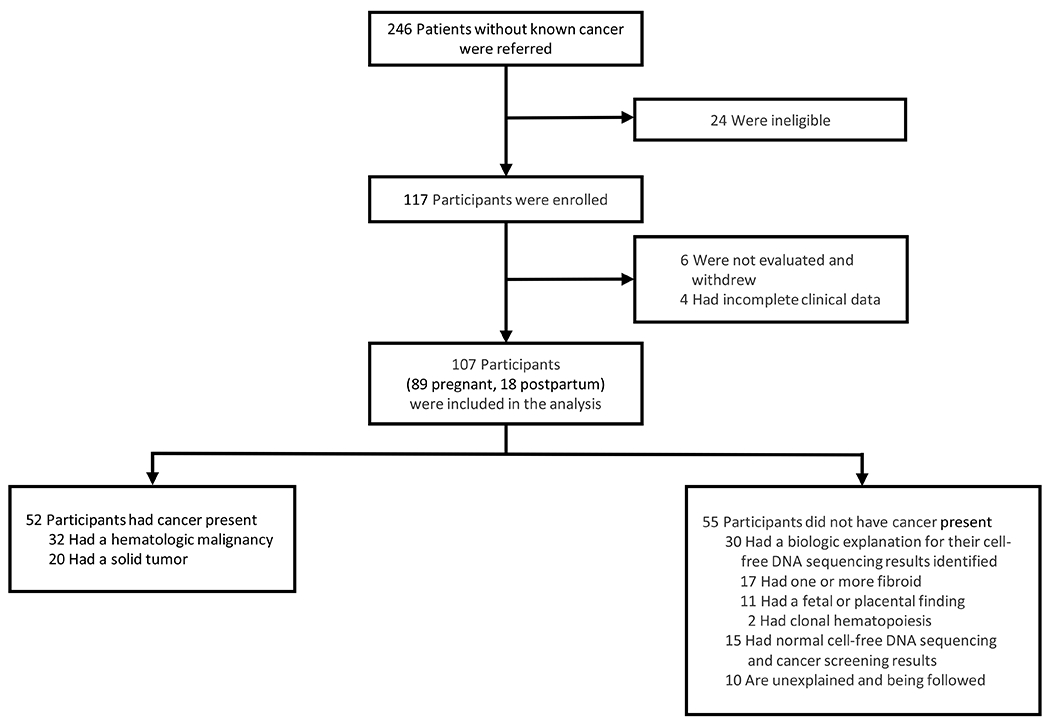
Methods: In this ongoing study we performed cancer screening in pregnant or postpartum persons who did not perceive signs or symptoms of cancer but received unusual clinical cfDNA-sequencing results or results that were nonreportable (i.e., the fetal aneuploidy status could not be assessed) from one of 12 different commercial laboratories in North America. We used a uniform cancer-screening protocol including rapid whole-body magnetic resonance imaging (MRI), laboratory tests, and standardized cfDNA sequencing for research purposes with the use of a genomewide platform. The primary outcome was the presence of cancer in participants after the initial cancer-screening evaluation. Secondary analyses included test performance.
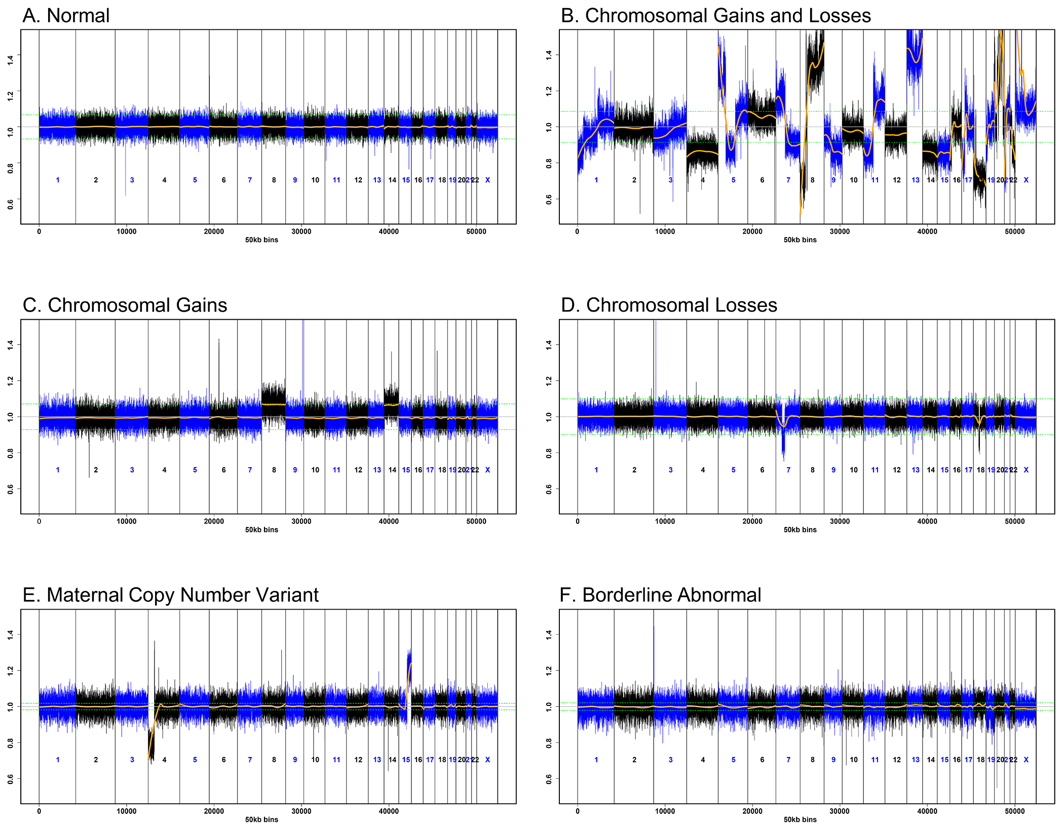
Results: Cancer was present in 52 of the 107 participants in the initial cohort (48.6%). The sensitivity and specificity of whole-body MRI in detecting occult cancer were 98.0% and 88.5%, respectively. Physical examination and laboratory tests were of limited use in identifying participants with cancer. Research sequencing showed that 49 participants had a combination of copy-number gains and losses across multiple (≥3) chromosomes; cancer was present in 47 of the participants (95.9%) with this sequencing pattern. Sequencing patterns of cfDNA in which there were only chromosomal gains (multiple trisomies) or only chromosomal losses (one or more monosomies) were found in participants with nonmalignant conditions, such as fibroids.
Conclusions: In this study, 48.6% of participants who received unusual or nonreportable clinical cfDNA-sequencing results had an occult cancer. Further study of DNA-sequencing patterns that are suggestive of occult cancer during prenatal screening is warranted. (Funded by the NIH Intramural Research Programs; ClinicalTrials.gov number, NCT04049604.).
Copyright © 2024 Massachusetts Medical Society.
Figures

Comment in
-
Prenatal cfDNA Sequencing and Incidental Detection of Maternal Cancer.N Engl J Med. 2025 Feb 27;392(9):932-933. doi: 10.1056/NEJMc2500437. N Engl J Med. 2025. PMID: 40009818 No abstract available.
-
Prenatal cfDNA Sequencing and Incidental Detection of Maternal Cancer. Reply.N Engl J Med. 2025 Feb 27;392(9):933. doi: 10.1056/NEJMc2500437. N Engl J Med. 2025. PMID: 40009819 No abstract available.
References
-
- Bianchi DW, Parker RL, Wentworth J, et al. DNA sequencing versus standard prenatal aneuploidy screening. N Engl J Med 2014;370:799–808. - PubMed
-
- Norton ME, Jacobsson B, Swamy GK, et al. Cell-free DNA analysis for noninvasive examination of trisomy. N Engl J Med 2015;372:1589–97. - PubMed
-
- American College of Obstetricians and Gynecologists’ Committee on Practice Bulletins—Obstetrics; Committee on Genetics; Society for Maternal-Fetal Medicine. Screening for fetal chromosomal abnormalities: ACOG Practice Bulletin, Number 226. Obstet Gynecol 2020;136:e48–e69. - PubMed
-
- Bianchi DW, Chudova D, Sehnert AJ, et al. Noninvasive prenatal testing and incidental detection of occult maternal malignancies. JAMA 2015;314:162–9. - PubMed
Publication types
MeSH terms
Substances
Associated data
Grants and funding
LinkOut - more resources
Full Text Sources
Medical