. 2024 Dec 30;19(12):e0315737.
doi: 10.1371/journal.pone.0315737. eCollection 2024.

Macro- and microscopic anatomy of the digestive tract in the red-eared slider (Emydidae: Trachemys scripta elegans)

Affiliations

Macro- and microscopic anatomy of the digestive tract in the red-eared slider (Emydidae: Trachemys scripta elegans)

Nonoha Miyai et al. PLoS One. .

Abstract

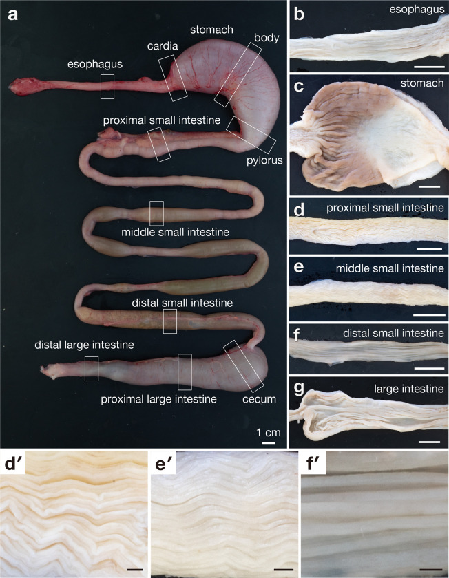
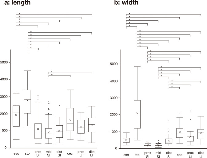
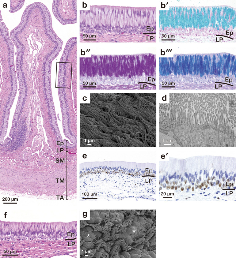
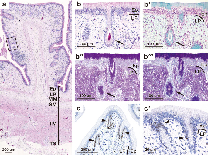
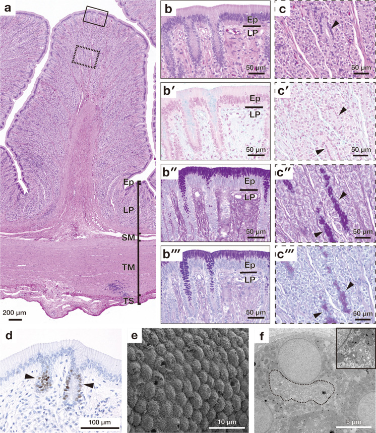
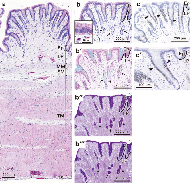
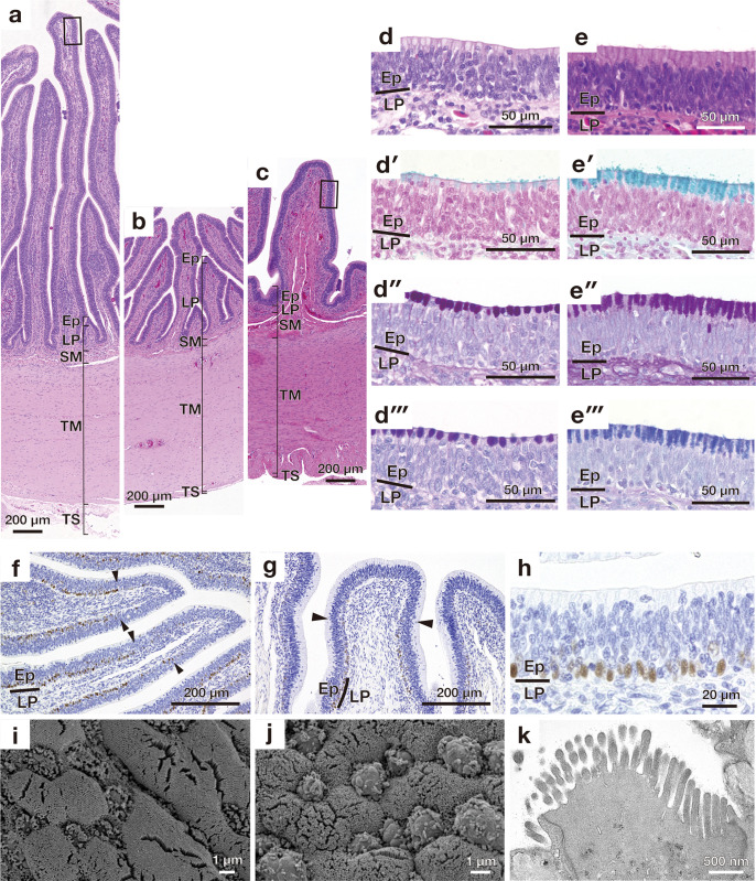
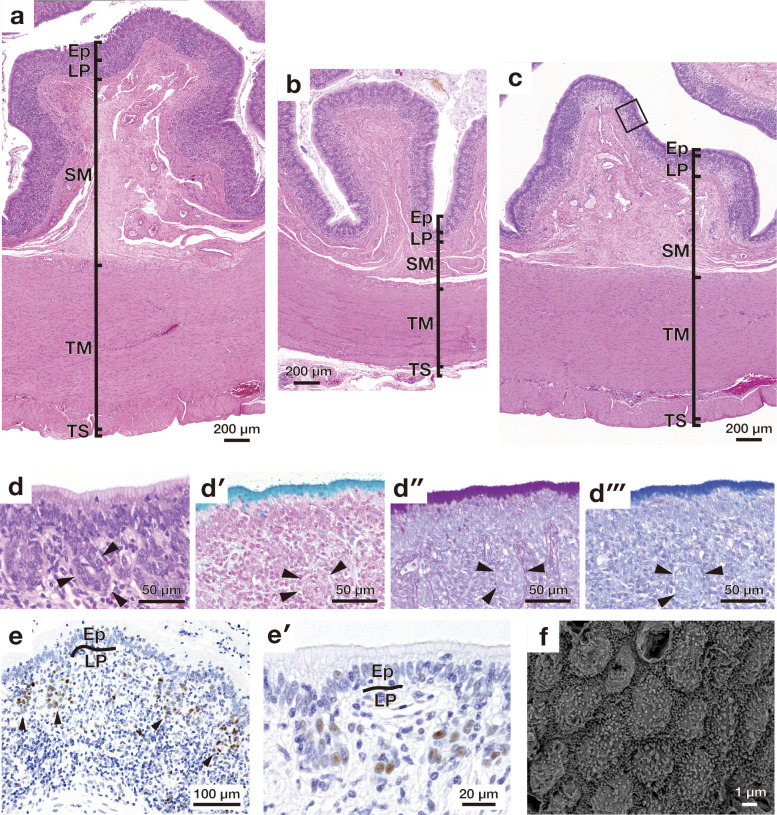
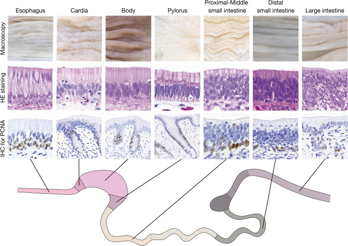
The red-eared sliders (Emydidae: Trachemys scripta) is characterised by a high adaptability to a variety of environment and threatens the habitat of Japanese native species. The ability to digest a variety of diets may attribute to the high adaptive capacity of this species to various environments, however, the digestive morphology remains scarcely described in red-eared sliders. In this study, we investigated the macro- and microscopic anatomy of the esophagus, stomach, small intestine, and large intestine in red-eared sliders. All segments of the digestive tract had longitudinal mucosal folds, the height and width of which varied in each segment of the digestive tract. The stomach had the highest and widest mucosal folds. The mucosal folds in the proximal-to-middle small intestine exhibited a zigzag shape, whereas those in the distal small intestine were linear. The wall of the digestive tract regularly consisted of mucosa, submucosa, tunica muscularis, and tunica adventitia or serosa. In each segment of the digestive tract, the epithelial structure was different. The esophagus and small intestine were lined by the pseudostratified columnar epithelium. In both segments, the basal part of the pseudostratified epithelium included proliferating cell nuclear antigen (PCNA)-positive proliferating cells. The stomach and large intestine were lined by the simple columnar epithelium. In the stomach and large intestine, PCNA-positive proliferating cells were present in the neck of the proper gastric gland and crypt-like structures, respectively. The proper gastric gland was composed of oxynticopeptic and mucous cells. This study revealed the detailed macro- and microscopic anatomy of the digestive tract in red-eared sliders. Overall, our findings may provide an anatomical basis for understanding the relationship between morphology and function in the digestive tract of turtles.


Conflict of interest statement

The authors declare that they have no known competing financial interest or personal relationships that could have appeared to influence the work reported in this paper.

Figures

Fig 1
Fig 1
Photographs of the unfolded digestive tube (a) and the mucosal folds of each segment of the digestive tract (b-g). (a) shows the unfolded digestive tract with blocks that indicate the sampling sites. (b)-(g) show the mucosal fold of the esophagus (b), stomach (c), proximal small intestine (d), middle small intestine (e), distal small intestine (f), and large intestine (g). (d′)-(f′) show the enlarged view of the mucosal folds if proximal, middle, and distal small intestine, respectively. Scale bar = 1 cm.
Fig 2
Fig 2
Box plots showing the statistical difference in the length (a) and width (b) of the mucosal fold in each segment of digestive tract. In each box, horizontal line indicates median value, box extend from 25% to 75% of distribution of values in each digestive segment, vertical extending lines indicate adjacent values, dots indicate outlier, and cross marks indicate mean value. *, P<0.01. eso, esophagus; sto, stomach; prox SI, proximal small intestine; mis SI, middle small intestine; dist SI, distal small intestine; cec, cecum; prox LI, proximal large intes tine; dist LI, distal large intestine.
Fig 3
Fig 3. Microscopic appearance of a mucosal fold in the esophagus and esophago-gastric junction.
(a) Overall cross-sectional view of a longitudinal fold stained with haematoxylin and eosin (HE). (b) shows a higher magnification of the mucosal epithelium enclosed by solid lines in (a). (b′)-(b‴) show high magnification images of the epithelial layer stained with alcian blue (AB) (b′), periodic acid Schiff (PAS) (b″), combined AB and PAS (b‴). (c) is scanning electron microscopy (SEM) image of the mucosal epithelium in the esophagus. (d) is transmission electron micrograph showing the cilia. (e) shows proliferating cell nuclear antigen-positive cells present in the basal part of the epithelium. (e′) shows a high magnification image of PCNA-positive cells. (f) shows cross-section of the epithelium of the esophago-gastric junction stained with HE. (g) SEM image of the mucosal epithelium in the esophago-gastric junction. Asterisks indicate the goblet cells. Ep, epithelium; LP, lamina propria; SM, submucosa; TM, tunica muscularis; TA, tunica adventitia.
Fig 4
Fig 4. Microscopic appearance of the gastric fold in the cardia.
(a) Overall cross-sectional view of the gastric fold stained with haematoxylin and eosin. (b) Higher magnification image of the mucosal epithelium enclosed by solid lines in (a). Arrow indicates the cardiac gland. (b′)-(b‴) show high magnification images of the epithelial layer stained with alcian blue (AB) (b′), periodic acid Schiff (PAS) (b″), combined AB and PAS (b‴). Arrow indicates the cardiac gland. (c) shows proliferating cell nuclear antigen (PCNA)-positive cells present in the basal part of the epithelium (indicated by arrowheads). (c′) shows a high magnification image of PCNA-positive cells. Arrowheads indicate PCNA positive cells. Ep, epithelium; LP, lamina propria; MM, muscularis mucosae; SM, submucosa; TM, tunica muscularis; TS, tunica serosa.
Fig 5
Fig 5. Microscopic appearance of the gastric fold in the body of stomach.
(a) Overall cross-sectional view of the gastric fold stained with haematoxylin and eosin. (b) Higher magnification image of the mucosal epithelium enclosed by solid lines in (a). (b′)-(b‴) show high magnification images of the epithelial layer stained with alcian blue (AB) (b′), periodic acid Schiff (PAS) (b″), combined AB and PAS (b‴). (c) Higher magnification image of the gastric gland enclosed by broken lines in (a). (c′)-(c‴) show high magnification images of the gastric gland stained with alcian blue (AB) (c′), periodic acid Schiff (PAS) (c″), combined AB and PAS (c‴). Arrowheads indicate the mucus cells in the gastric gland. (d) shows proliferating cell nuclear antigen-positive cells present in the neck of the gastric gland. (e) Scanning electron micrograph of the mucosal epithelium. (f) Transmission electron micrograph of an oxynticopeptic cell. The secretory canaliculus locates in the area enclosed by the broken line. Inset shows a high magnification view of the secretory granule (indicated by asterisk) and mitochondria. Ep, epithelium; LP, lamina propria; SM, submucosa; TM, tunica muscularis; TS, tunica serosa.
Fig 6
Fig 6. Microscopic appearance of the gastric fold in the pylorus.
(a) Overall cross-sectional view of the gastric fold stained with haematoxylin and eosin. (b) Higher magnification image of the mucosal fold. Arrows indicate the pyloric gland. Inset shows higher magnification image of the epithelium of the mucosal fold. (b′)-(b‴) show high magnification images of the mucosal fold stained with alcian blue (AB) (b′), periodic acid Schiff (PAS) (b″), combined AB and PAS (b‴). Arrows indicate the pyloric gland. (c) shows proliferating cell nuclear antigen (PCNA)-positive cells present in the basal part of the epithelium (indicated by arrowheads). (c′) shows a high magnification image of PCNA-positive cells. Arrowheads indicate PCNA positive cells. Ep, epithelium; LP, lamina propria; MM, muscularis mucosae; SM, submucosa; TM, tunica muscularis; TS, tunica serosa.
Fig 7
Fig 7. Microscopic appearances of longitudinal folds in the small intestine.
Overall cross-sectional view of mucosa stained with haematoxylin and eosin in the proximal (a), middle (b), and distal (c) small intestines. (d) Higher magnification image of the mucosal epithelium enclosed by solid lines in (a). (d′)-(d‴) show high magnification images of the epithelial layer stained with alcian blue (AB) (d′), periodic acid Schiff (PAS) (d″), combined AB and PAS (d‴). (e) Higher magnification image of the mucosal epithelium enclosed by solid lines in (c). (e′)-(e‴) show high magnification images of the epithelial layer stained with alcian blue (AB) (e′), periodic acid Schiff (PAS) (e″), combined AB and PAS (e‴). (f) and (g) show proliferating cell nuclear antigen (PCNA)-positive cells present in the basal part of the epithelium of the proximal (f) and distal (g) small intestine. Arrowheads indicate the boundary between the apical and middle portions of the mucous fold. (h) shows a high magnification image of PCNA-positive cells in the epithelium. (i) and (j) are scanning electron microscopy images of the mucosal epithelium of the proximal (i) and distal (j) small intestine. (k) Transmission electron micrograph of enterocyte having microvilli. Ep, epithelium; LP, lamina propria; SM, submucosa; TM, tunica muscularis; TS, tunica serosa.
Fig 8
Fig 8. Microscopic appearance of mucosal folds in the large intestine.
(a)-(c) show the overall cross-sectional views of mucosa stained with haematoxylin and eosin in the cecum (a), proximal large intestine (b), and distal large intestine (c). (d) Higher magnification image of the mucosal epithelium enclosed by solid lines in (c). Arrowheads indicate the crypt-like structure. (d′)-(d‴) show high magnification images of the epithelial layer stained with alcian blue (AB) (d′), periodic acid Schiff (PAS) (d″), combined AB and PAS (d‴). Arrowheads indicate the crypt-like structure. (e) shows proliferating cell nuclear antigen (PCNA)-positive cells present in the crypt-like structure. (e′) shows a high magnification image of PCNA-positive cells. (f) Scanning electron micrograph of the mucosal epithelium of the large intestine. Ep, epithelium; LP, lamina propria; SM, submucosa; TM, tunica muscularis; TS, tunica serosa.
Fig 9
Fig 9. Graphical summary of the findings of the present study.
Upper, middle, and lower lines show the macroscopic morphology of the mucosal folds, micrographs of epithelium stained with hematoxylin an eosin, and micrographs of proliferating cell nuclear antigen-positive cell in each segment of digestive tract, respectively. HE, hematoxylin an eosin; IHC, immunohistochemistry; and PCNA, proliferating cell nuclear antibody.

Similar articles

References

    1. Crawford NG, Parham JF, Sellas AB, Faircloth BC, Glenn TC, Papenfuss TJ, et al.. A phylogenomic analysis of turtles. Mol Phylogenet Evol. 2015;83: 250–257. doi: 10.1016/j.ympev.2014.10.021 - DOI - PubMed
    1. Rasmussen AR, Murphy JC, Ompi M, Gibbons JW, Uetz P. Marine reptiles. PLOS One. 2011;6: e27373. doi: 10.1371/journal.pone.0027373 - DOI - PMC - PubMed
    1. Chatterji RM, Hipsley CA, Sherratt E, Hutchinson MN, Jones EH. Ontogenetic allometry underlies trophic diversity in sea turtles (Chelonioidea). Evol Ecol. 2022;36: 511–540. doi: 10.1007/s10682-022-10162-z - DOI
    1. Shipps BK, Peecook BR, Angielczyk KD. The topography of diet: orientation patch count predicts diet in turtles. Anat Rec. 2023;306: 1214–1227. doi: 10.1002/ar.25125 - DOI - PubMed
    1. Hofmann RR. Evolutionary steps of ecophysiological adaptation and diversification of ruminants: a comparative view of their digestive system. Oecologia. 1989;78: 443–457. doi: 10.1007/BF00378733 - DOI - PubMed

MeSH terms

LinkOut - more resources