. 2025 Jan 7;20(1):e0317035.
doi: 10.1371/journal.pone.0317035. eCollection 2025.

Evidence of avian and human influenza A virus infection in farmed Siamese crocodiles (Crocodylus siamensis) in Thailand

Affiliations

Evidence of avian and human influenza A virus infection in farmed Siamese crocodiles (Crocodylus siamensis) in Thailand

Metawee Thongdee et al. PLoS One. .

Abstract

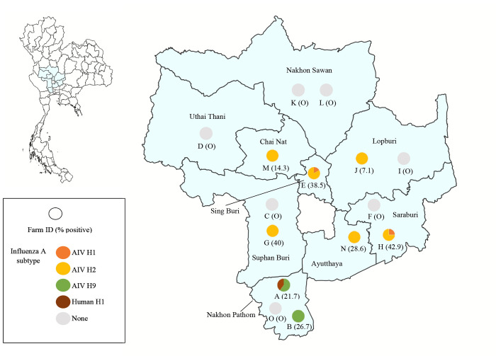
Crocodilians are susceptible to a range of virus infection including influenza A virus (IAV). However, little is known about the ecology and epidemiology of IAV in crocodile species. This study aimed to investigate IAV infection in farmed Siamese crocodiles in central Thailand. We collected plasma samples and pharyngeal swab samples from Siamese crocodiles residing in 13 crocodile farms in 9 provinces of central Thailand during 2019. Additional archival plasma samples of Siamese crocodiles collected in 2012 and 2018 were also included in the study. Plasma samples were screened for influenza A antibodies by a hemagglutination inhibition (HI) assay and positive were evaluated by a cytopathic effect/hemagglutination based-microneutralization (MN) assay. Swab samples were tested for influenza viral RNA by a real-time RT-PCR assay targeting the influenza matrix (M) gene. Among 246 tested plasma samples, the overall seroprevalence of antibodies against IAV in farmed Siamese crocodiles was 17.5% (43/246). The most common hemagglutinin (HA) subtype was H2 (46.5%, 20/43) followed by H9 (39.5%, 17/43), human H1 (14%, 6/43) and H1 (7%, 3/43). Multiple HA subtypes were also detected in 7% (3/43) of infected crocodiles with combination of H1 and H2 subtypes. All 126 tested swab samples were negative for influenza viral RNA. In addition, we demonstrated the ability of wild-type IAV subtypes (H1, H2, H9 and human H1) to infect primary Siamese crocodile fibroblast cells. To our knowledge, this is the first report of serological evidences of avian and human IAV infection in Siamese crocodiles. Our findings highlighted the role of crocodile species in the ecology of IAV particularly the potential to serve as the reservoir or mixing vessel for the viruses that significantly threaten both human and animal health.


Conflict of interest statement

The authors have declared that no competing interests exist.

Figures

Fig 1
Fig 1. Map of nine provinces of central Thailand and distribution of seropositive cases against influenza A subtypes in plasma of farmed Siamese crocodiles by HI and MN assay.
The circle represents the farm location and the positivity observed with the identified influenza A subtypes. The map was generated using QGIS (Quantum Geographic Information System) version 3.34.13 (https://qgis.org). Geographical materials used for creating the map (e.g., shapefiles) were supported by the DIVA-GIS database (https://www.diva-gis.org/). Source of base map: GADM (https://gadm.org/license.html).
Fig 2
Fig 2. Western blot analysis showing the presence of crocodile IgY antibody in the infected crocodile plasma.
Crocodile plasma samples (No. 1 and No. 2) at 1:50, 1:100 and 1:200 dilutions were run on SDS-PAGE gels for protein separation. The blotted membranes were probed with HRP conjugated chicken IgY antibody and protein bands were visualized after adding chromogenic substrate. (A) On non-reducing 8% SDS-PAGE gel, the crocodile IgY molecule (Y) was observed at MW of approximately 180 kDa (B) On reducing 12% SDS-PAGE gel, crocodile IgY heavy chain and light chain were observed at MW of approximately 60–70 kDa and 25 kDa, respectively. Chicken serum samples at 1:50 and 1:100 dilutions were included as control which the chicken IgY were detected with HRP conjugated-goat anti-chicken IgY antibody. A standard protein marker with the indicated MW (in kDa) was included in each SDS-PAGE gel. Raw western blot images are available in S1 Fig.
Fig 3
Fig 3. Immunofluorescence staining of primary crocodile fibroblast cells at 24 hours post-infection with AIV H1N1, AIV H2N8, AIV H9N2 and human H1N1 (TH104).
Mock-infected cells were included as negative control. The influenza virus nucleoprotein (NP) localization was observed using Alexa fluor 488-conjugated mouse antibody (in green). Cell nucleus DNA was counterstained with DAPI (in blue). Magnification, X200.

Similar articles

References

    1. Long JS, Mistry B, Haslam SM, Barclay WS. Host and viral determinants of influenza A virus species specificity. Nat Rev Microbiol. 2019; 17:67–81. doi: 10.1038/s41579-018-0115-z - DOI - PubMed
    1. Tong S, Li Y, Rivailler P, Conrardy C, Castillo DA, Chen LM, et al.. A distinct lineage of influenza A virus from bats. Proc Natl Acad Sci U S A. 2012; 109(11):4269–74. doi: 10.1073/pnas.1116200109 - DOI - PMC - PubMed
    1. Tong S, Zhu X, Li Y, Shi M, Zhang J, Bourgeois M, et al.. New world bats harbor diverse influenza A viruses. PLoS Pathog. 2013; 9(10):e1003657. doi: 10.1371/journal.ppat.1003657 - DOI - PMC - PubMed
    1. Webster RG, Bean WJ, Gorman OT, Chambers TM, Kawaoka Y. Evolution and ecology of influenza A viruses. Microbiol Rev. 1992; 56(1):152–79. doi: 10.1128/mr.56.1.152-179.1992 - DOI - PMC - PubMed
    1. Medina RA, García-Sastre A. Influenza A viruses: New research developments. Nat Rev Microbiol. 2011; 9(8):590–603. doi: 10.1038/nrmicro2613 - DOI - PMC - PubMed

MeSH terms

Substances

LinkOut - more resources