. 2025 Jan 2;66(1):17.
doi: 10.1167/iovs.66.1.17.

Idebenone Protects Photoreceptors Impaired by Oxidative Phosphorylation Disorder in Retinal Detachment

Affiliations

Idebenone Protects Photoreceptors Impaired by Oxidative Phosphorylation Disorder in Retinal Detachment

Lisong Wang et al. Invest Ophthalmol Vis Sci. .

Abstract

Purpose: Oxidative phosphorylation (OXPHOS) is an aerobic metabolic mechanism, and its dysfunction plays an important role in the pathological changes of ischemic diseases. However, systematic studies on the occurrence of retinal detachment (RD) are lacking.

Methods: Single-cell RNA sequencing (scRNA-seq) of the human retina was performed to detect the metabolic changes of various retinal cells after RD. In this study, animal experiments were conducted to explore the OXPHOS activity after RD. In addition, idebenone, a coenzyme Q10 (CoQ10) analog currently used to treat Leber hereditary optic neuropathy (LHON), was used to improve the OXPHOS disorder in experimental RD model.

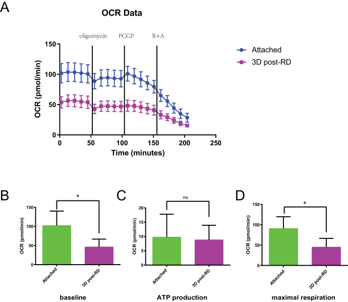
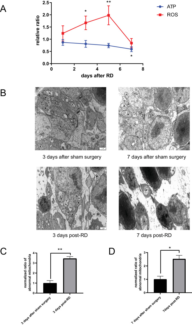
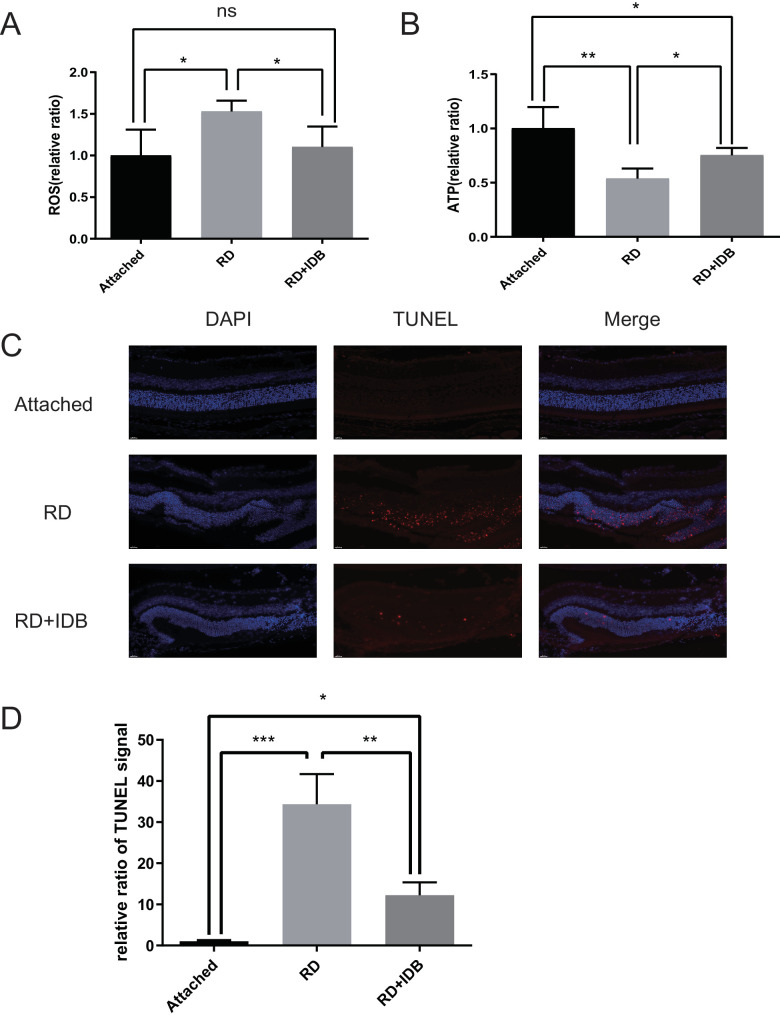
Results: ScRNA-seq revealed abnormal energy metabolism and OXPHOS pathways in retinal cells after RD. Adenosine triphosphate (ATP) and reactive oxygen species (ROS) are the main products of OXPHOS, the mouse RD model indicated that the rise in ROS levels may have a greater impact on photoreceptors in the early stage, whereas decreased ATP synthesis was observed in the later stage; these changes threaten the function and morphology of the retina. Idebenone was administered to model mice intragastrically, leading to reduced ROS levels in the early stage post-RD and improved ATP synthesis in the later stage, which was closely related to the maintenance of mitochondrial morphology.

Conclusions: OXPHOS disorder leads to photoreceptor degeneration after RD, which can be alleviated by improving OXPHOS function.


Conflict of interest statement

Disclosure: L. Wang, None; G. Zou, None; Y. Yan, None; R. Shi, None; Y. Guo, None; M. Zhang, None; L. Lu, None; K. Dong, None

Figures

Figure 1.
Figure 1.
The OCR of the retinal tissues of mice decreased after RD. (A) OCR time series curves of the attached group (n = 6) and three days post-RD (n = 6). (B) Baseline OCR at day 3 (55.11 ± 23.93, P = 0.031) post-RD was significantly lower than that of the attached group (102.32 ± 37.75). (C) ATP production at three days (8.82 ± 5.10) post-RD was not significantly different from that of attached group (9.79 ± 7.96). (D) The maximal respiration at three days (46.29 ± 20.55, P = 0.018) post-RD was significantly lower than that of the attached group (92.53 ± 32.63).
Figure 2.
Figure 2.
Abnormal mitochondrial morphology and function were observed in photoreceptors in the experimental RD model. (A) Time series curves of ROS and ATP post-RD: ROS increased significantly at day 3 (1.67 ± 0.28, P = 0.021, n = 4) and day 5 (1.98 ± 0.39, P = 0.007, n = 4) post-RD compared with the attached group. ATP had no significant change on day 1 (0.87 ± 0.12, n = 4), day 3 (0.81 ± 0.14, n = 4) and day 5 (0.74 ± 0.09, n = 4) post-RD, and was significantly lower than that of attached group on day 7 (0.59 ± 0.10, P = 0.04, n = 4) post-RD. (BD) Morphological changes of mitochondria post-RD: on the third (3.44 ± 0.35, P = 0.002, n = 3) and seventh (2.52 ± 0.47, P = 0.013, n = 3) day post-RD, the mitochondria became vacuolar and the proportion of irregular mitochondria increased significantly compared with the attached group (n = 3). indicates abnormal mitochondria; * indicates normal mitochondria.
Figure 3.
Figure 3.
OXPHOS dysfunction in the experimental RD model. (AF) Changes of OXPHOS complex activity after RD (n = 3): The activities of NDUFB8 (complex I) decreased significantly on the seventh day (0.54 ± 0.14, P = 0.007) post RD, and SDHB (complex II) decreased significantly on the fifth day (0.46 ± 0.14, P = 0.003) and seventh day (0.37 ± 0.12, P < 0.001) post RD; UQCRFS1 (complex III), COX4 (complex IV), and ATP5A (complex V) had no significant changes compared with control group in whole time series.
Figure 4.
Figure 4.
Idebenone preserved OXPHOS function post-RD. (A) IDB mitigated the rise of ROS at day 3 post-RD: the ROS levels in the IDB group (1.10 ± 0.24, n = 3) were significantly lower than that in the RD group (1.52 ± 0.13, P = 0.036, n = 3). (B) IDB mitigated ATP decline on day 7 after RD: ATP content was significantly higher in the IDB group (0.75 ± 0.07, n = 4) compared to the RD group (0.54 ± 0.09, P = 0.045, n = 4). (C) IDB reduced photoreceptor apoptosis on day 3 post-RD: the apoptotic signal was significantly reduced in the IDB group(12.24 ± 3.10, n = 3) compared with the RD group (34.35 ± 7.32, P = 0.001, n = 3).

Similar articles

References

    1. Govers BM, van Huet RAC, Roosing S, et al. .. The genetics and disease mechanisms of rhegmatogenous retinal detachment. Prog Retin Eye Res. 2023; 97: 101158. - PubMed
    1. Qureshi MH, Steel DHW.. Retinal detachment following cataract phacoemulsification-a review of the literature. Eye (Lond). 2020; 34: 616–631. - PMC - PubMed
    1. Melo IM, Zhou TE, Nagel F, et al. .. Histological changes in retinal detachment: a systematic review for the clinician. Surv Ophthalmol. 2024; 69: 85–92. - PubMed
    1. Yan Y, Wang Y, Ding J, Lu L, Ke GJ, Dong K.. TRPML1 inhibited photoreceptor apoptosis and protected the retina by activation of autophagy in experimental retinal detachment. Ophthalmic Res. 2021; 64: 587–594. - PubMed
    1. Jiang L, Yin X, Chen YH, et al. .. Proteomic analysis reveals ginsenoside Rb1 attenuates myocardial ischemia/reperfusion injury through inhibiting ROS production from mitochondrial complex I. Theranostics. 2021; 11: 1703–1720. - PMC - PubMed

MeSH terms