. 2025 Jan 7;15(1):1054.
doi: 10.1038/s41598-024-82921-7.

Ovario- protective effect of Moringa oleifera leaf extract against cyclophosphamide-induced oxidative ovarian damage and reproductive dysfunction in female rats

Affiliations

Ovario- protective effect of Moringa oleifera leaf extract against cyclophosphamide-induced oxidative ovarian damage and reproductive dysfunction in female rats

Seham Samir Soliman et al. Sci Rep. .

Abstract

It is crucial to develop new tactics to prevent ovarian tissue damage in women whose reproductive toxicity is caused by chemotherapy. The present investigation was performed to assess the protective effects of Moringa oleifera (M. oleifera) leaf extract on cyclophosphamide (CP)-induced ovarian damage and reproductive dysfunction. Thirty-two female, healthy Wistar albino rats were randomly assigned to four groups (8 rats/group). The first group was given saline intraperitoneally (i.p.). The second group was given a single dose of CP (200 mg/kg; i.p.). The third and fourth groups were given M. oleifera leaf extract (150 and 250 mg/kg; orally) for 20 days before receiving CP on the final day of the experiment. Hormonal assessments, including follicle stimulating hormone (FSH), luteinizing hormone (LH), and estrogen (ES), were performed 24 h after CP administration. In addition, the antioxidant status and inflammatory response against CP were evaluated. Moreover, detailed histopathological and ultra- structural observations were conducted. For evaluation of statistical significance between different groups; One-way analysis of variance (ANOVA) with Tukey's post hoc test was adopted. Our findings revealed that rats subjected to CP showed increased levels of FSH, LH, malondialdehyde (MDA), tumor necrosis factor-alpha, and interleukin-8 and decreased levels of ES and glutathione. Pre-treatment with M. oleifera leaf extract (250 mg/kg; orally) was statistically significant (p values < 0.05) as it could improve hormonal changes, oxidative stress indices, and pro- inflammatory mediator levels. Consequently, a marked improvement was observed in the ovarian and uterine architectures, with a normal ovarian reserve and a normal endothelium with normal tubular glands. In conclusion, M. oleifera leaf extract (250 mg/kg) could be used as a pharmaceutical supplement because it protects female rats against CP-induced ovarian damage and reproductive dysfunction.

Keywords: Moringa oleifera; Cyclophosphamide; Ovarian function; Oxidative stress; Pro-inflammatory mediators.


Conflict of interest statement

Declarations. Competing interests: The authors declare no competing interests. Ethics approval and consent to participate: All experimental procedures were conducted following the recommendations of the National Institutes of Health Guide for Care and Use of Laboratory Animals (NIH Publications No. 8023, revised 1978) and after the approval of the National Research Centre–Medical Research Ethics Committee (NRC-MREC) for the use of animal subjects (Approval No. 2411022023).

Figures

Fig. 1
Fig. 1
Effect of administration of M. oleifera leaf extract on the photographs of the genital tract of female rats received CP induced reproductive dysfunction. Reproductive dysfunction was induced in female rats by i.p. administration of CP (200 mg/kg. b.w.). Rats were treated with M. oleifera leaf extract (150 and 250 mg/kg; orally). At the end of the study, rats were sacrificed by cervical dislocation and their genital tract was weighted and photographed. Where (a; control, b; CP group, c; CP + M. oleifera (150 mg/kg) group, d; CP + M. oleifera (250 mg/kg) group.
Fig. 2
Fig. 2
Effect of administration of M. oleifera leaf extract on the FSH and LH values in female rats received CP induced reproductive dysfunction. Reproductive dysfunction was induced in female rats by i.p. administration of CP (200 mg/kg. b.w.). Rats were treated with M. oleifera leaf extract (150 and 250 mg/kg; orally). 24 h after the last dose of CP, serum levels of FSH and LH were evaluated. Results are expressed as mean ± SD (n = 8). *Significant difference from control group, p < 0.05. @ Significant difference from CP- received group, P < 0.05.
Fig. 3
Fig. 3
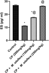
Effect of administration of M. oleifera leaf extract on the ES values in female rats received CP induced reproductive dysfunction. Reproductive dysfunction was induced in female rats by i.p. administration of CP (200 mg/kg. b.w.). Rats were treated with M. oleifera leaf extract (150 and 250 mg/kg; orally). 24 h after the last dose of CP, serum levels of ES were evaluated. Results are expressed as mean ± SD (n = 8). *Significant difference from control group, p < 0.05. @ Significant difference from CP- received group, P < 0.05.
Fig. 4
Fig. 4
Effect of administration of M. oleifera leaf extract on GSH and MDA values in female rats received CP induced reproductive dysfunction. Reproductive dysfunction was induced in female rats by i.p. administration of CP (200 mg/kg. b.w.). Rats were treated with M. oleifera leaf extract (150 and 250 mg/kg; orally). 24 h after the last dose of CP, ovarian levels of GSH and MDA were evaluated. Results are expressed as mean ± SD (n = 8). *Significant difference from control group, p < 0.05. @ Significant difference from CP- received group, P < 0.05.
Fig. 5
Fig. 5
Effect of administration of M. oleifera leaf extract on TNF-α and IL-8 values in female rats received CYP induced reproductive dysfunction. Reproductive dysfunction was induced in female rats by i.p. administration of CP (200 mg/kg. b.w.). Rats were treated with M. oleifera leaf extract (150 and 250 mg/kg; orally). 24 h after the last dose of CP, ovarian levels of TNF-α and IL-8 were evaluated. Results are expressed as mean ± SD (n = 8). *Significant difference from control group, p < 0.05. @ Significant difference from CP- received group, P < 0.05.
Fig. 6
Fig. 6
Photomicrographs of H&E staining of ovarian sections of control and different experimental groups, (A,B) Control group. (A) showed normal ovarian histoarchitecture, normal growing follicles till normal preovulatory follicles (PF), normal Medulla (MD) and corpus luteum (CL) can also be seen.(B) Showed normal preovulatory follicle are seen with large follicular antrum (FA) and corona radiate (CR) surrounding the oocyte (OC). A normal cortical stroma can be seen with the theca folliculi (TF) surrounding the granulosa cells (GC). (C,D) Cyclophosphamide group. (C) exhibited apoptotic signs are seen on the antral follicles as in degenerating (atretic) tertiary follicle (AF) of with distorted granulosa cells (GC) and in damage preovulatory follicle (PF), completely atretic secondary follicle (SF), with the appearance fibrotic corpus luteum (CL). (D) Preovulatory follicle show vacuolated oocyte surrounded by fibrotic granulosa cells (GC). (E,F) CP + M. oleifera 150 mg/kg group showed mild damage of the ovarian tissue, primordial follicle (PMF) appear normal, the secondary follicle (SF) with normal size and abnormal shape. (F) Preovulatory follicle (PF) still with few signs of apoptosis as unhealthy oocyte (OC) and vacuolated (V) follicular antrum (FA). (G,H) CP + M. oleifera 250 mg/kg (G) showed most ovarian tissue preserves its normal histological appearance with normal growing cells as: primordial follicle (PMF), secondary follicle (SF) and Preovulatory follicle (PF), with presence of few vacuolated follicles (arrow). (H) Nearly normal Preovulatory follicle with typical structure. (Magnification: (A,C,E,G) 10X and (B,D,F,H) 40X).
Fig. 7
Fig. 7
Photomicrographs of H&E staining of uterus sections of control and different experimental groups, (A) group Control group showed normal uterine architecture presented with intact endothelium (E) , normal tubular glands (TG) and patent wide uterine Lumen (UL) with different layers; endometrium (End), myometrium (myo) with normal thickness (double head arrows). (B) CP (200 mg/kg) group exhibited noticed uterine section degeneration, appear of massive leukocyte infiltration (Yellow Arrow), congested tubular glands (TG), multi vacuoles (V), and endothelium with apoptotic epithelium cells. (C) CP + M. oleifera 150 mg/kg group showi mildly disrupted uterine appearance, endometrium (End) without infiltration, few epithelial vacuolar degeneration (V) and slight Congested tubular gland (TG). (D) CP + M. oleifera 250 mg/kg showed marked Improvement of uterine section architecture, normal endothelium (E), normal tubular glands (TG) and few vacuoles (V) in endometrium (End). (Magnification 40X).
Fig. 8
Fig. 8
TEM micrograph of ovary from control and different treatment groups: (A,B) control group (A) showing a single oocyte follicle (SOFs) with a clear round shaped nucleus (N) and normal mitochondria (M) distributed throughout the cytoplasm and oriented as a grape like structure.(B) showing Normal ovarian interstitial cells (IC) with contacted cell–cell junction (arrow). (C,D) CP (200 mg/kg) group exhibited degeneration of primordial follicle follicles (Pf), follicles nucleus appeared shrinked losing its normal shape and structure, with appearing of multi-vaculation (V). Follicles reach to attritic phase (*), with gapping between ovarian cells (white arrow). (E,F) CP + M. oleifera 150 mg/kg group showing slight improvement of single oocyte follicle , where its nucleus preserve its normal round cells (NN) and other nucleus appear irregular (IN).with still appearing of little multivaculation (V) .(G,H) CP + M. oleifera 250 mg/kg showed ovarian tissue appear near normal to control group where the primordial follicles with normal oval shape and its nucleus appear nearly normal (N) with its normal distributed chromatin (Ch) and normal mitochondria (M).

References

    1. Siegel, R. L. et al. Cancer statistics, 2023. Ca Cancer J. Clin.73 (1), 17–48 (2023). - PubMed
    1. Elmariah, H. et al. Haploidentical bone marrow transplantation with post-transplant cyclophosphamide using non–first-degree related donors. Biol. Blood Marrow Transpl.24 (5), 1099–1102 (2018). - PMC - PubMed
    1. Song, D. et al. Human umbilical cord mesenchymal stem cells therapy in cyclophosphamide-induced premature ovarian failure rat model. Biomed. Res. Int.2016, 1–13 (2016). - PMC - PubMed
    1. Wang, H. et al. Inflammation activates the interferon signaling pathways in taste bud cells. J. Neurosci.27 (40), 10703–10713 (2007). - PMC - PubMed
    1. Wang, X. et al. Efficacy of pegfilgrastim to support neoadjuvant dose-dense epirubicin and cyclophosphamide chemotherapy in breast cancer. Support. Care Cancer27, 3019–3025 (2019). - PubMed

Publication types

LinkOut - more resources