

Kinematics of balance controls in people with chronic ankle instability during unilateral stance on a moving platform
- PMID: 39774772
- PMCID: PMC11707225
- DOI: 10.1038/s41598-025-85220-x
Kinematics of balance controls in people with chronic ankle instability during unilateral stance on a moving platform
Abstract
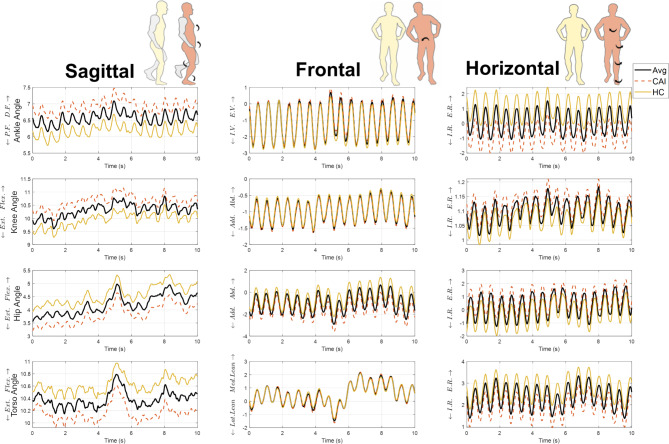
Balance control deficits resulting from ankle sprains are central to chronic ankle instability (CAI) and its persistent symptoms. This study aimed to identify differences in balance control between individuals with CAI and healthy controls (HC) using challenging single-leg balance tasks. Twenty-three CAI and 23 HC participants performed balance tasks on a force plate that either remained static or moved mediolaterally. Force and kinematic data were recorded to measure balance and joint movements. The CAI group showed significantly shorter time-to-boundary during static conditions but no significant differences during moving conditions compared to HC. During moving conditions, CAIs exhibited greater proximal compensations, with greater range of motion and higher angular velocity in the knee, hip, and torso. while no significant differences were observed in these parameters during static conditions. Principal component analysis indicated specific kinetic chain in CAI during one-leg balance under both static and moving conditions compared to HC. These findings suggest an altered movement strategy in CAI, that ankle injuries impair the ability to stabilize both distal and proximal joints, and an altered kinetic chain from ankle to torso. Rehabilitation programs for CAI might benefit from considering the integration of the entire kinetic chain, addressing both distal and proximal joint dynamics to support effective recovery and prevent secondary injuries.
Keywords: Balance control; Chronic ankle instability; Kinematics; Perturbation; Postural strategy; Principal component analysis.
© 2025. The Author(s).
Conflict of interest statement
Declarations. Competing interests: The authors declare no competing interests.
Figures


Similar articles
-
Trunk-rotation differences at maximal reach of the star excursion balance test in participants with chronic ankle instability.J Athl Train. 2015 Apr;50(4):358-65. doi: 10.4085/1062-6050-49.3.74. Epub 2014 Dec 22. J Athl Train. 2015. PMID: 25531142 Free PMC article.
-
Lower Limb Joint Kinetics During a Side-Cutting Task in Participants With or Without Chronic Ankle Instability.J Athl Train. 2020 Feb;55(2):169-175. doi: 10.4085/1062-6050-334-18. Epub 2020 Jan 2. J Athl Train. 2020. PMID: 31895591 Free PMC article.
-
Lower Extremity Biomechanics During a Drop-Vertical Jump in Participants With or Without Chronic Ankle Instability.J Athl Train. 2018 Apr;53(4):364-371. doi: 10.4085/1062-6050-481-15. Epub 2018 Apr 18. J Athl Train. 2018. PMID: 29667844 Free PMC article.
-
Proximal Adaptations in Chronic Ankle Instability: Systematic Review and Meta-analysis.Med Sci Sports Exerc. 2020 Jul;52(7):1563-1575. doi: 10.1249/MSS.0000000000002282. Med Sci Sports Exerc. 2020. PMID: 31977639
-
Treatment of common deficits associated with chronic ankle instability.Sports Med. 2009;39(3):207-24. doi: 10.2165/00007256-200939030-00003. Sports Med. 2009. PMID: 19290676 Review.
Cited by
-
Differences in lower extremity kinematics and kinetics during a side-cutting task in patients with and without chronic ankle instability.Front Sports Act Living. 2025 Jun 6;7:1593231. doi: 10.3389/fspor.2025.1593231. eCollection 2025. Front Sports Act Living. 2025. PMID: 40546779 Free PMC article.
-
Identifying the Most Sensitive COP Variable in Static Balance: The Impact of Chronic Ankle Instability on Postural Stability.J Foot Ankle Res. 2025 Sep;18(3):e70072. doi: 10.1002/jfa2.70072. J Foot Ankle Res. 2025. PMID: 40750356 Free PMC article.
-
Humans self-organise balance control strategies on a dynamic platform.Sci Rep. 2025 Jul 8;15(1):24366. doi: 10.1038/s41598-025-09127-3. Sci Rep. 2025. PMID: 40628838 Free PMC article.
References
-
- Delahunt, E. et al. Clinical assessment of acute lateral ankle sprain injuries (ROAST): 2019 consensus statement and recommendations of the International Ankle Consortium. Br. J. Sports Med.52(20), 1304–1310. 10.1136/bjsports-2017-098885 (2018). - PubMed
-
- Hiller, C. E. et al. Prevalence and impact of chronic musculoskeletal ankle disorders in the community. Arch. Phys. Med. Rehabil.93(10), 1801–1807. 10.1016/j.apmr.2012.04.023 (2012). - PubMed
-
- Yu, P., Mei, Q., Xiang, L., Fernandez, J. & Gu, Y. Differences in the locomotion biomechanics and dynamic postural control between individuals with chronic ankle instability and copers: A systematic review. Sports Biomech.21(4), 531–549. 10.1080/14763141.2021.1954237 (2022). - PubMed
Publication types
MeSH terms
Grants and funding
LinkOut - more resources
Full Text Sources
Medical
