. 2025 Feb;32(2):393-404.
doi: 10.1038/s41594-024-01452-x. Epub 2025 Jan 7.

Structural basis for the inhibition of PRC2 by active transcription histone posttranslational modifications

Affiliations

Structural basis for the inhibition of PRC2 by active transcription histone posttranslational modifications

Trinity Cookis et al. Nat Struct Mol Biol. 2025 Feb.

Abstract

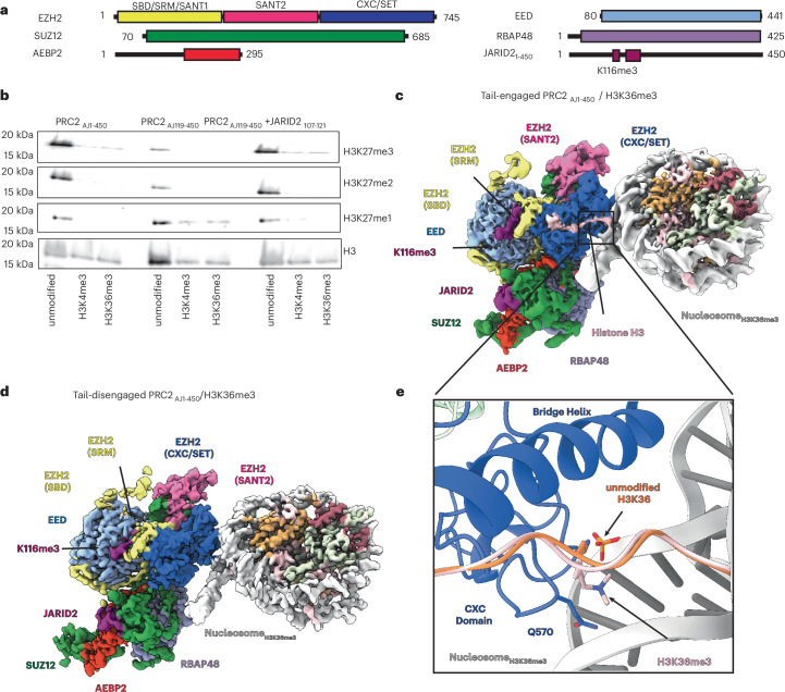
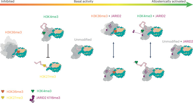
Polycomb repressive complex 2 (PRC2) trimethylates histone H3 on K27 (H3K27me3) leading to gene silencing that is essential for embryonic development and maintenance of cell identity. PRC2 is regulated by protein cofactors and their crosstalk with histone modifications. Trimethylated histone H3 on K4 (H3K4me3) and K36 (H3K36me3) localize to sites of active transcription and inhibit PRC2 activity through unknown mechanisms. Using cryo-electron microscopy, we reveal that histone H3 tails containing H3K36me3 engage poorly with PRC2 and preclude its effective interaction with chromatin, while H3K4me3 binds to the allosteric site in the EED subunit, acting as an antagonist that competes with activators required for spreading of the H3K27me3 repressive mark. Thus, the location of the H3K4me3 and H3K36me3 modifications along the H3 tail allows them to target two requirements for efficient trimethylation of H3K27 by PRC2. We further show that the JARID2 cofactor modulates PRC2 activity in the presence of these histone modifications.


Conflict of interest statement

Competing interests: The authors declare no competing interests.

Figures

Fig. 1
Fig. 1. Cryo-EM structures of PRC2AJ1450 bound to H3K36me3-modified nucleosomes.
a, Schematic representation of protein domains in the PRC2–AEBP2–JARID2 complex used in this work, containing either JARID21450 or JARID2119. b, Representative methyltransferase assays performed on mononucleosome substrates that were either unmodified, H3K4me3 modified or H3K36me3 modified. Assays were repeated in triplicate with PRC2AJ1450, PRC2AJ119450 or PRC2AJ119450 in the presence of 150 µM methylated JARID2 peptide including residues 107–121. c, Cryo-EM structure of PRC2AJ1450 bound to an H3K36me3-modified nucleosome in which the H3 tail is engaged by EZH2 and PRC2 is in an allosterically stimulated state. d, Cryo-EM structure of PRC2AJ1450 bound to H3K36me3-modified nucleosomes in which the H3 tail is not engaged by EZH2. The structures shown in c,d coexist in the sample. e, Comparison of the position of K36 with respect to the nucleosomal DNA in structures of PRC2 bound to unmodified H3K36 (shown in orange; PDB 6WKR) and H3K36me3-modified nucleosomes (shown in pink). Source data
Fig. 2
Fig. 2. Comparison of tail-engaged and tail-disengaged PRC22AJ1450 complexes bound to H3K36me3-modified nucleosomes.
a, Overlay of the cryo-EM density maps for the coexisting tail-engaged (blue) and tail-disengaged (green) PRC2AJ1450–H3K36me3 structures identified by our analysis. Maps are aligned using the nucleosome to show the relative rotation of PRC2 on the nucleosome surface. b, Close-up view of the EZH2 bridge helix showing its relative position with respect to the H3 tail (pink) and nucleosomal DNA (gray) for the tail-engaged (blue) and tail-disengaged (green) structures. c, Close-up view of the EZH2 CXC domain showing its relative position with respect to the H3 tail and nucleosomal DNA for the tail-engaged (blue) and tail-disengaged (green) structures (in b,c, the tail, as seen in the tail-engaged complex, is shown in pink).
Fig. 3
Fig. 3. Cryo-EM structures of PRC2 bound to H3K4me3-modified nucleosomes.
a, Cryo-EM structure of PRC2AJ119450 bound to H3K4me3-modified nucleosomes. b, Close-up view of the cryo-EM density showing the N terminus of histone H3 containing H3K4me3 bound to EED. The map is shown at a threshold of 0.487 (lower threshold in Extended Data Fig. 7a). c, Cryo-EM structure of PRC2AJ1450 bound to H3K4me3-modified nucleosomes. d, Close-up view of cryo-EM density showing JARID2K116me3 bound to EED and the folding of the SRM helix. The map is shown at a threshold of 0.25.
Fig. 4
Fig. 4. H3K4me3 acts as an allosteric antagonist by binding to EED.
a, Interactions between the region of histone H3 (pink) around the H3K4me3 modification and the aromatic cage of EED (light blue). b, Interactions involving JARID2 (magenta), EED (light blue) and EZH2 (yellow for the SRM and SAL; darker blue for the SET domain) in an allosterically activated PRC2. The coordinates used were from the PRC2AJ1450–H3K4me3 structure obtained in this study (Fig. 3c). JARID2 R115 interacts with E137 and D140 of the EZH2 SRM. JARID2 R108 and R110 are positioned to interact with E162 and D160 of EZH2, while JARID2 L111 and EZH2 I150 are involved in hydrophobic contacts. c, Interactions between the peptide around H3K27me3 (pink) and EED (light blue) and EZH2 (yellow for the SRM and SAL; darker blue for the SET domain). The coordinates used are those from the X-ray crystal structure of an activated PRC2 catalytic lobe from Chaetomium thermophilum (PDB 5KJH). Histone H3R26 (similarly to JARID2 R115) interacts with negatively charged residues in the EZH2 SRM and histone H3R23 establishes additional contacts with the SRM. d, Sequence alignment with respect to the PRC2-methylated lysine for the N-terminal region of the histone H3 tail around K4, JARID2 around K116, H3 around K27 and PALI1 around K1241. The PRC2-modified lysine is colored in red, residues that are involved in hydrophobic contacts are colored in tan and residues involved in electrostatic interactions are colored in blue. e, Overlay of structures shown in a (colored) and b (gray or transparent) showing that histone H3R2 of H3K4me3 clashes with the SAL, with the region corresponding to peptides bound to the allosteric site zoomed in on the right.
Fig. 5
Fig. 5. Model for the regulation of PRC2 catalytic activity by modifications of the H3 tail in the nucleosome substrate and the presence or absence of methylated JARID2.
Trimethylation of histone H3K27 by PRC2 can be inhibited by H3K36me3 and H3K4me3 histone modifications and activated by H3K27me3 (in trans) or JARID2 K116me3. The interplay between these regulators is a gradient of PRC2 activity (red to green arrow). From left to right: in the absence of JARID2, H3K36me3 prevents efficient tail engagement and results in the loss of a well-defined register between PRC2 and the nucleosome substrate, depicted as the blurry model of PRC2; histone H3K4me3 engages the allosteric site and competes with nucleosomes that are already modified with H3K27me3; PRC2 on unmodified substrates in the absence of JARID2 has a basal level of activity, while its EED allosteric site remains open for possible engagement with nearby nucleosomes with H3K27me3; in the presence of JARID2, PRC2 is allosterically stimulated, resulting in increased H3K27me3 activity. Histone H3K36me3 and H3K4me3 reduce PRC2 activity through the same mechanisms described above; however, methylated JARID2 stabilizes the catalytic lobe of PRC2, facilitates some tail engagement in the presence of H3K36me3 and can compete with H3K4me3 for the EED allosteric site.
Extended Data Fig. 1
Extended Data Fig. 1. Processing workflow for PRC2AJ1-450 bound to H3K36me3 nucleosomes.
Data collected for PRC2AJ1-450 bound to H3K36me3 nucleosomes was initially processed in RELION. Focused classification around the nucleosome and EZH2 SET domain resulted in two classes, one showing the histone H3 tail engaged and one lacking density for the histone H3 tail. We further refined particles with the tail engaged and performed focused classification around the PRC2 top lobe to obtain a 3.1 Å reconstruction that resolved the histone H3K36me3 side chain (left side of processing diagram). We then used the initial 4,011,989 particles obtained from 2D classification to perform 3D classification and heterogeneous refinement to select particles lacking density for the histone H3 tail followed by homogeneous and local refinement using CryoSPARC to resolve the 3.5 Å reconstruction of the ‘tail-disengaged’ state (right side of processing diagram).
Extended Data Fig. 2
Extended Data Fig. 2. Local resolution mapped onto cryo-EM maps of PRC2AJ1-450 bound to H3K36me3-containing nucleosomes obtained from local refinements using masks surrounding PRC2 and nucleosome regions.
For each map, the resolution at FSC = 0.143 is provided, while the map-to-model FSC plots show the masked resolution at FSC = 0.5. Each map has been locally filtered using the local resolution estimate. Angular distribution plot for particles that make up the final reconstructions are also included. A) PRC2AJ1-450 / H3K36me3 ‘tail-engaged’ state B) Examples of fitting of protein amino acid side chains into the density map for each subunit within the PRC2 complex. C) PRC2AJ1-450 / H3K36me3 ‘tail-disengaged’ state D) Examples of fitting of protein amino acid side chains into the density map for each subunit within the PRC2 complex E) Close up view of active site density observed in the PRC2AJ1-450 / H3K36me3 ‘tail-disengaged’ state corresponding to either JARID2 or the histone H3 tail. JARID2 residues 23-29 (magenta) are shown docked into the residual density but were excluded from the deposited coordinates. F) Cryo-EM density shown for the histone H3 tail (pink) bound to the EZH2 SET domain (blue) in the PRC2AJ1-450 / H3K36me3 ‘tail-engaged’ state. G) Close up view of histone H3K27 (pink) bound to the active site of EZH2 (blue).
Extended Data Fig. 3
Extended Data Fig. 3. Cryo-EM of PRC2AJ119-450 bound to H3K36me3, unmodified, and H3Δ38 nucleosomes.
A) Cryo-EM analysis of PRC2AJ119-450 bound to H3K36me3 modified nucleosomes. Data was extensively cleaned by 2D classification trying to bring up density for PRC2. Representative 2D classes after 6 rounds are shown, but the resulting 214,262 particles resulted in a 4.6 Å reconstruction showing only the nucleosome, without additional density for PRC2. Further cleaning of the data by 2D classification (after 13 total rounds) only showed fuzzy density for PRC2. B) Data collected for PRC2AJ119-450 bound to unmodified nucleosomes was processed in cryosparc following standard workflow. Particles were subjected to 2D classification, followed by heterogeneous refinement to remove damaged complexes. Non-uniform refinement was performed followed by local refinement around PRC2. A second dataset of 441 movies was collected and used to generate a reconstruction showing density of PRC2 for comparison with H3K36me3 and H3Δ38 complexes. C) Cryo-EM of PRC2AJ119-450 bound to nucleosomes containing H3Δ38. Data was extensively cleaned by 2D classification trying to bring up density for PRC2. Representative 2D classes after 13 rounds are shown. The resulting 24,172 particles were used to obtain a 6.4 Å reconstruction that shows only the nucleosome, without additional density for PRC2, despite the weak density that can be observed in the 2D classification.
Extended Data Fig. 4
Extended Data Fig. 4. Cryo-EM and nucleosome binding analysis of PRC2 with H3K36me3, unmodified, and H3Δ38 nucleosomes.
A) Representative 2D classes of PRC2AJ119-450 bound to H3K36me3 nucleosomes show absence of strong density for PRC2, despite observed binding in electromobility shift assays. Representative electromobility shift assays were performed with a titration of 0 to 400 nM PRC2 with 50 nM nucleosome in all cases. Results were reproduced in triplicate. B) Representative 2D classes of PRC2AJ119-450 bound to nucleosomes containing histone H3Δ38 show absence of strong density for PRC2, despite observed binding in electromobility shift assays. C) Representative 2D classes of PRC2AJ119-450 bound to unmodified nucleosomes that yielded the 4.5 Å reconstruction. D) Representative 2D classes of PRC2AJ1-450 bound to H3K36me3 nucleosomes that yielded the 3.6 Å tail-engaged reconstruction. In contrast to A and B, C and D show strong density for PRC2. Electromobility shift assays show similar binding affinity for PRC2AJ119-450 bound to unmodified nucleosomes or PRC2AJ1-450 bound to H3K36me3 nucleosomes to that observed for the PRC2AJ119-450/NucleosomeH3K36me3 and PRC2AJ119-450/NucleosomeH3Δ38 assays.
Extended Data Fig. 5
Extended Data Fig. 5. Processing workflow for PRC2AJ119-450 bound to H3K4me3 nucleosomes.
Data collected for PRC2AJ119-450 bound to H3K4me3 nucleosomes was initially processed in cryosparc. Two data sets were merged after 2D classification and one round of heterogeneous refinement. Particles were imported into RELION for 3D Refinement followed by 3D classification without alignment. All 3D classes show density for the histone H3 tail and absence of density for the EZH2 SRM. Classes were selected showing the strongest density for H3K4me3 bound to EED. After CTF Refinement, particles were imported into cryosparc for non-uniform refinement and local refinements around the nucleosome and PRC2 regions. Source data
Extended Data Fig. 6
Extended Data Fig. 6. Local resolution maps for PRC2AJ119-450 complexes bound to H3K4me3-containing nucleosomes.
A) Local resolution mapped onto cryo-EM maps of PRC2AJ119-450 complexes bound to H3K4me3-containing nucleosomes obtained from local refinements using masks surrounding PRC2 or nucleosome regions. For each map, the resolution at FSC = 0.143 is provided, while the map-to-model FSC plots show the masked resolution at FSC = 0.5. Each map has been locally filtered using the local resolution estimate. Angular distribution plot for particles that make up the final reconstructions are also included B) Examples of fitting of protein amino acid side chains into the density map for each subunit within the PRC2 complex. C) Cryo-EM density shown for the histone H3 tail (pink) bound to the EZH2 SET domain (blue). D) Close up view of histone H3K27 (pink) bound to the active site of EZH2 (blue).
Extended Data Fig. 7
Extended Data Fig. 7. Comparison of cryo-EM density for the EED allosteric site in structures of PRC2AJ119-450 bound to H3K4me3 or to unmodified nucleosomes.
A) Density surrounding the H3K4me3 bound to the EED for the PRC2AJ119-450 bound to a H3K4me3-modified nucleosome shown at threshold of 0.232 B) Cryo-EM density map (transparent grey) and model of the EED allosteric site and the bound methylated peptide for PRC2AJ119-450 bound to a H3K4me3-modified nucleosome. The model is shown in ribbon representation colored by subunit, with EED shown in light blue and the histone H3K4me3 peptide shown in pink. C) Corresponding view of a reconstruction obtained for PRC2AJ119-450 bound to a unmodified nucleosome showing lack of density in the EED allosteric pocket. D) Corresponding view of our recently reported reconstruction obtained for PRC2AJ119-450 bound to a an unmodified nucleosome that additionally contained the linker histone H1, also showing lack of density in the EED allosteric pocket.
Extended Data Fig. 8
Extended Data Fig. 8. Processing workflow for PRC2AJ1-450 bound to H3K4me3 nucleosomes.
Data collected for PRC2AJ1-450 bound to H3K4me3 nucleosomes was processed in cryosparc. Two data sets were merged after 2D classification and heterogeneous refinement. Particles were imported into RELION and subjected to 3D classification without alignment. Focused classification was performed using a mask around the allosteric site and EZH2 SRM regions resulting in two major classes showing the presence of absence of the SRM. Each class was refined separately and imported into cryosparc for local refinement around the PRC2 region. Close up view of the allosteric site of the PRC2AJ1-450 / H3K4me3 cryo-EM map (grey) showing the corresponding SRM density (yellow) in the ‘activated’ PRC2AJ1-450 / H3K4me3 cryo-EM map that has been lowpass filtered to to 8 Å. Both maps contain an unassigned density marked with asterisk that is not apparent in any of the other cryo-EM density maps of PRC2/nucleosome complexes.
Extended Data Fig. 9
Extended Data Fig. 9. Local resolution maps for PRC2AJ1-450 complexes bound to H3K4me3-containing nucleosomes.
A) Local resolution mapped onto cryo-EM maps of PRC2AJ1-450 complexes bound to H3K4me3-containing nucleosomes obtained from local refinements using masks surrounding PRC2 or nucleosome regions. For each map, the resolution at FSC = 0.143 is provided, while the map-to-model FSC plots show the masked resolution at FSC = 0.5. Each map has been locally filtered using the local resolution estimate. PRC2AJ1-450 / H3K4me3 B) Examples of fitting of protein amino acid side chains into the density map for each subunit within the PRC2 complex C) Cryo-EM density shown for the histone H3 tail (pink) bound to the EZH2 SET domain (blue). D) Close up view of histone H3K27 (pink) bound to the active site of EZH2 (blue).
Extended Data Fig. 10
Extended Data Fig. 10. Activation of PRC2AJ119-450 by methylated JARID2 peptides.
A) Methylated JARID2 peptide sequences used to perform methyltranserfase assays. B) Representative methyltransferase assays performed with PRC2AJ119-450 complexes on unmodified mononucleosome substrates in the absence or in the presence of 15 µM (+) or 150 µM (++) methylated JARID2 peptides. Results were reproduced in triplicate. C) Cryo-EM density for PRC2AJ119-450/unmodified nucleosome complex in the presence of JARID2107-121 shows strong density for the SRM helix. Map displayed after lowpass filtering to 7 Å. D) Cryo-EM density for PRC2AJ119-450/unmodified nucleosome complex in the presence of JARID2113-121,R115A shows density corresponding to the peptide, but weak density for the SRM. Map displayed is lowpass filtered to 7 Å. Source data

Update of

References

    1. Cao, R. et al. Role of histone H3 lysine 27 methylation in Polycomb-group silencing. Science298, 1039–1043 (2002). - PubMed
    1. Hauri, S. et al. A high-density map for navigating the human Polycomb complexome. Cell Rep17, 583–595 (2016). - PubMed
    1. Grijzenhout, A. et al. Functional analysis of AEBP2, a PRC2 Polycomb protein, reveals a Trithorax phenotype in embryonic development and in ESCs. Development143, 2716–2723 (2016). - PMC - PubMed
    1. Poepsel, S., Kasinath, V. & Nogales, E. Cryo-EM structures of PRC2 simultaneously engaged with two functionally distinct nucleosomes. Nat. Struct. Mol. Biol.25, 154–162 (2018). - PMC - PubMed
    1. Kasinath, V. et al. JARID2 and AEBP2 regulate PRC2 in the presence of H2AK119ub1 and other histone modifications. Science371, eabc3393 (2021). - PMC - PubMed