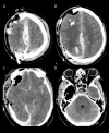
Malignant Cerebral Edema After Cranioplasty: A Case Report and Literature Insights
- PMID: 39774935
- PMCID: PMC11725662
- DOI: 10.12659/AJCR.946230
Malignant Cerebral Edema After Cranioplasty: A Case Report and Literature Insights
Abstract
BACKGROUND Decompressive craniectomy is a common life-saving intervention in the setting of elevated intracranial pressure. Cranioplasty restores the calvarium and intracranial physiology once swelling recedes. Cranioplasty is often thought of as a low-risk intervention. However, numerous reports indicate that malignant cerebral edema (MCE) is an often-fatal complication of an otherwise uneventful cranioplasty. A careful review of the literature is needed to better understand this devastating condition. CASE REPORT A 41-year-old man presented after suffering a gunshot wound to the right frontal lobe. Upon initial evaluation, the patient had grossly visible brain matter, left-sided hemiparesis with a Glascow Coma Score (GCS) of 11, and vital signs concerning for elevated intracranial pressure. Computed tomography (CT) showed right-sided intraparenchymal and subarachnoid hemorrhage with a 5 mm leftward midline shift. The patient was taken to the operating room (OR) for right fronto-parietal craniectomy. Over the next 3 months, he recovered steadily and underwent PEEK cranioplasty on post-operative day 83. Pre-operative CT showed sunken skin flap syndrome with an 8-mm midline shift. Following an uneventful cranioplasty, he failed to regain consciousness. Examination revealed absent brainstem reflexes. CT showed global diffuse cerebral edema. The patient was declared brain dead. CONCLUSIONS Continued research is needed to better understand the pathophysiology of malignant cerebral edema so that future incidences may be prevented. A combination of negative-pressure suction drainage, sunken skin flap syndrome, and delayed time to cranioplasty likely play a significant role in the evolution of MCE. We urge neurosurgeons to consider the likelihood of MCE and adapt surgical planning accordingly.
Conflict of interest statement
Figures


References
-
- Diaz-Segarra N, Jasey N. Improved rehabilitation efficiency after cranioplasty in patients with sunken skin flap syndrome: A case series. Brain Inj. 2024;38(2):61–67. - PubMed
-
- Lilja-Cyron A, Andresen M, Kelsen J, et al. Intracranial pressure before and after cranioplasty: Insights into intracranial physiology. J Neurosurg. 2020;133(5):1548–58. - PubMed
-
- Zanaty M, Chalouhi N, Starke RM, et al. Complications following cranioplasty: Incidence and predictors in 348 cases. J Neurosurg. 2015;123(1):182–88. - PubMed
Publication types
MeSH terms
LinkOut - more resources
Full Text Sources

