. 2025 Jan 8;15(1):1249.
doi: 10.1038/s41598-025-85138-4.

Ten-year outcomes after DMEK, DSAEK, and PK: insights on graft survival, endothelial cell density loss, rejection and visual acuity

Affiliations

Ten-year outcomes after DMEK, DSAEK, and PK: insights on graft survival, endothelial cell density loss, rejection and visual acuity

Theresa Isabelle Wilhelm et al. Sci Rep. .

Abstract

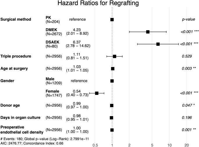
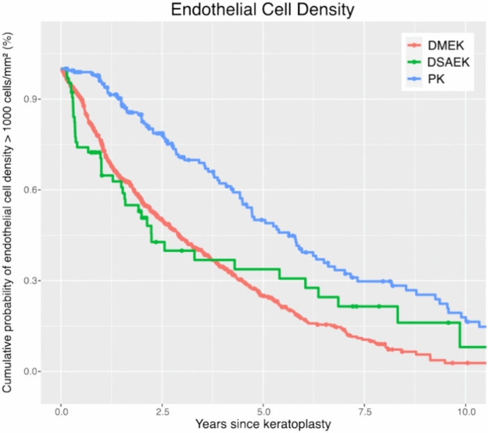
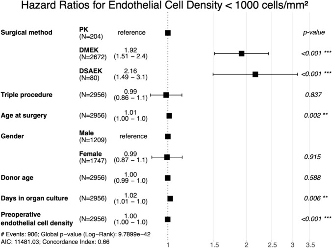
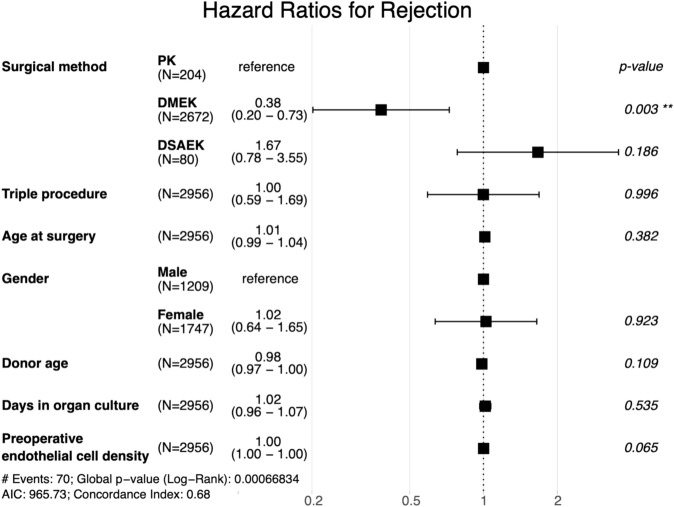
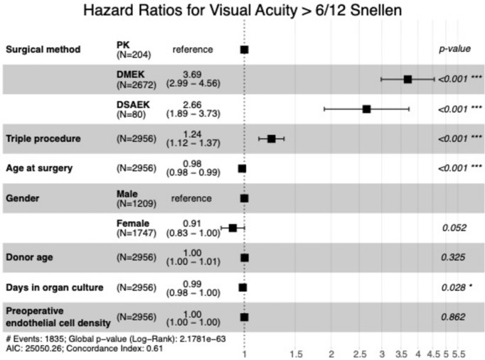
Fuchs Endothelial Corneal Dystrophy (FECD) is the most frequent indication for corneal transplantation, with Descemet membrane endothelial keratoplasty (DMEK), Descemet stripping automated endothelial keratoplasty (DSAEK), and penetrating keratoplasty (PK) being viable options. This retrospective study compared 10-year outcomes of these techniques in a large cohort of 2956 first-time keratoplasty eyes treated for FECD at a high-volume corneal transplant center in Germany. While DMEK and DSAEK provided faster visual recovery (median time to BSCVA ≥ 6/12 Snellen: DMEK 7.8 months, DSAEK 12.4 months, PK 37.9 months; cumulative probability of BSCVA ≥ 6/12 Snellen within 5 years: DMEK 93%, DSAEK 83%, PK 63%), PK surprisingly exhibited superior long-term graft survival (92% vs. 75% for DMEK and 73% for DSAEK at 10 years). Endothelial cell density (ECD) decreased significantly faster after DMEK and DSAEK, potentially contributing to their lower graft survival (10-year ECD > 1000 cells/mm2 probability: DMEK 3%, DSAEK 8%, PK 18%). DMEK demonstrated the lowest rejection rate (10% at 10 years vs. 13% for PK and 19% for DSAEK). These findings challenge the perceived superiority of endothelial keratoplasty and highlight the need for further investigation into the long-term implications of accelerated endothelial cell loss after DMEK and DSAEK.

Keywords: Corneal transplantation; DMEK; DSAEK; Fuchs endothelial corneal dystrophy; Graft survival; Penetrating keratoplasty.


Conflict of interest statement

Declarations. Competing interests: The authors declare no competing interests. Generative AI and AI-assisted technologies in the writing process: Grammarly v1.72.3.0, DeepL, GPT-4o, and Claude 3.5 Sonnet were used for linguistic improvements. The authors carefully checked the text suggestions for accuracy and made corrections where necessary. They take full responsibility for the publication’s content.

Figures

Fig. 1
Fig. 1
Flowchart of study inclusion criteria and resulting patient cohort.
Fig. 2
Fig. 2
Graft survival: Kaplan–Meier cumulative probability of regraft-free survival within ten years after DMEK, DSAEK, and PK. The survival rate is higher after PK compared to DMEK and DSAEK. DMEK Descemet membrane endothelial keratoplasty, DSAEK Descemet stripping automated endothelial keratoplasty, PK penetrating keratoplasty.
Fig. 3
Fig. 3
Hazard ratios for regrafting: Forest plot depicting adjusted hazard ratios with 95% confidence intervals for regrafting within ten years after PK, DMEK, and DSAEK. Hazard ratios are adjusted for clinical and demographic covariates, as listed on the left side of the plot. Triple procedure refers to a combination of keratoplasty with phacoemulsification and intraocular lens implantation. The probability of regrafting is higher after DMEK and DSAEK compared to PK. DMEK Descemet membrane endothelial keratoplasty, DSAEK Descemet stripping automated endothelial keratoplasty, PK penetrating keratoplasty.
Fig. 4
Fig. 4
Endothelial cell density loss: Kaplan–Meier cumulative probability of ECD dropping below 1000 cells/mm2 within ten years after DMEK, DSAEK, and PK. ECD drops faster following DMEK and DSAEK compared to PK. ECD Endothelial cell density, DMEK Descemet membrane endothelial keratoplasty, DSAEK Descemet stripping automated endothelial keratoplasty, PK penetrating keratoplasty.
Fig. 5
Fig. 5
Hazard Ratios for Endothelial Cell Density < 1000 cells/mm2: Forest plot depicting adjusted hazard ratios with 95% confidence intervals for endothelial cell density dropping below 1000 cells/mm2 within ten years after PK, DMEK, and DSAEK. Hazard ratios are adjusted for clinical and demographic covariates, as listed on the left side of the plot. Triple procedure refers to a combination of keratoplasty with phacoemulsification and intraocular lens implantation. The risk of endothelial cell density being lower than 1000 cells/mm2 is higher after DMEK and DSAEK compared to PK. DMEK Descemet membrane endothelial keratoplasty, DSAEK Descemet stripping automated endothelial keratoplasty, PK penetrating keratoplasty.
Fig. 6
Fig. 6
Rejection-free survival: Kaplan–Meier cumulative probability of rejection-free survival within ten years after DMEK, DSAEK, and PK. Higher rejection rates are observed after PK compared to DMEK. DMEK Descemet membrane endothelial keratoplasty, DSAEK Descemet stripping automated endothelial keratoplasty, PK penetrating keratoplasty.
Fig. 7
Fig. 7
Hazard ratios for rejection: Forest plot depicting adjusted hazard ratios with 95% confidence intervals for rejection within ten years after PK, DMEK, and DSAEK. Hazard ratios are adjusted for clinical and demographic covariates, as listed on the left side of the plot. Triple procedure refers to a combination of keratoplasty with phacoemulsification and intraocular lens implantation. The risk of rejection is lower after DMEK compared to PK. DMEK Descemet membrane endothelial keratoplasty, DSAEK Descemet stripping automated endothelial keratoplasty, PK penetrating keratoplasty.
Fig. 8
Fig. 8
Visual acuity rehabilitation: Kaplan–Meier cumulative probability of visual rehabilitation following keratoplasty (DMEK, DSAEK, and PK). BSCVA of 6/12 is achieved faster following DMEK and DSAEK compared to PK. DMEK Descemet membrane endothelial keratoplasty, DSAEK Descemet stripping automated endothelial keratoplasty, PK penetrating keratoplasty.
Fig. 9
Fig. 9
Hazard Ratios for Visual Acuity > 6/12 Snellen: Forest plot depicting adjusted hazard ratios with 95% confidence intervals for reaching a vision better than BSCVA 6/12 Snellen within ten years after PK, DMEK, and DSAEK. Hazard ratios are adjusted for clinical and demographic covariates, as listed on the left side of the plot. Triple procedure refers to a combination of keratoplasty with phacoemulsification and intraocular lens implantation. The probability of reaching a visual acuity above 6/12 Snellen is higher after DMEK and DSAEK compared to PK. DMEK Descemet membrane endothelial keratoplasty, DSAEK Descemet stripping automated endothelial keratoplasty, PK penetrating keratoplasty.

Similar articles

Cited by

References

    1. Ong Tone, S. et al. Fuchs endothelial corneal dystrophy: The vicious cycle of fuchs pathogenesis. Prog. Retin. Eye Res.80, 100863. 10.1016/j.preteyeres.2020.100863 (2021). - PMC - PubMed
    1. Gain, P. et al. Global survey of corneal transplantation and eye banking. JAMA Ophthalmol.134, 167. 10.1001/jamaophthalmol.2015.4776 (2016). - PubMed
    1. Altamirano, F. et al. fuchs endothelial corneal dystrophy: An updated review. Int. Ophthalmol.44, 61. 10.1007/s10792-024-02994-1 (2024). - PubMed
    1. Melles, G. R. J., Ong, T. S., Ververs, B. & van der Wees, J. Descemet membrane endothelial keratoplasty (DMEK). Cornea25, 987–990. 10.1097/01.ico.0000248385.16896.34 (2006). - PubMed
    1. Sarnicola, C., Farooq, A. V. & Colby, K. Fuchs endothelial corneal dystrophy: Update on pathogenesis and future directions. Eye Contact Lens Sci. Clin. Pract.45, 1–10. 10.1097/ICL.0000000000000469 (2019). - PubMed

LinkOut - more resources