Review
. 2025 Jul;50(7):2858-2867.
doi: 10.1007/s00261-024-04769-9. Epub 2025 Jan 7.

Standardizing the reporting of cholangiocarcinoma: the society of abdominal radiology disease focused panel on cholangiocarinoma lexicon

Affiliations
Review

Standardizing the reporting of cholangiocarcinoma: the society of abdominal radiology disease focused panel on cholangiocarinoma lexicon

Robert M Marks et al. Abdom Radiol (NY). 2025 Jul.

Abstract

In March 2023, the Society of Abdominal Radiology (SAR) Disease Focused Panel (DFP) on Cholangiocarcinoma (CCA) was formed. One of its initial tasks was for creation of a lexicon specific for CCA to complement the terms related to the Liver Imaging Reporting and Data System (LI-RADS) category M. A committee was formed and vetted 15 unique terms for CCA. The multidisciplinary members of the DFP passed each term by over 90% approval. The purpose of this paper is to describe the process for developing the lexicon, introduce the lexicon terms, and provide a pictorial atlas of the 15 vetted terms relating to the imaging findings of CCA.

Keywords: Cholangiocarcinoma; Lexicon; Liver cancer.


Conflict of interest statement

Declarations. Competing interests: The authors declare no competing interests.

Figures

Fig. 1
Fig. 1
Intraductal CCA. Axial CT images with contrast demonstrate an intraductal perihilar mass (arrow) with upstream intrahepatic ductal dilatation (A). There is tumor thrombus in the left portal vein (curved arrow) (B)
Fig. 2
Fig. 2
Mass Forming CCA. Axial T2 weighted image (A), and post-contrast T1 weighted arterial phase (B) and delayed phase (C) images demonstrate a large intrahepatic mass forming CCA that is mildly T2 bright (thick arrow, A), has rim APHE (arrow B) and delayed central enhancement and peripheral washout (thin arrow, C). There is associated capsular retraction (curved arrow)
Fig. 3
Fig. 3
Periductal Infiltrating Cholangiocarcinoma.Coronal magnetic resonance cholangiopancreatography (MRCP) demonstrates a stricture of the common bile duct (CBD) (A). Incidentally noted is a 2 cm side branch intraductal papillary mucinous neoplasm in the pancreatic head (curved arrow). Post contrast T1 weighted image demonstrates narrowing of the CBD with wall thickening and enhancement (B)
Fig. 4
Fig. 4
Intrahepatic Cholangiocarcinoma. Axial T2 (A), and post contrast T1-weighted portal venous phase (B) and delayed phase (C) images demonstrate a 6.1 cm mass forming CCA (arrow) in hepatic segment 7. There is rim enhancement on the portal venous phase (B) with centripetal central enhancement on the delayed phase (C). There are dilated upstream bile ducts peripheral to this mass (curved arrows)
Fig. 5
Fig. 5
Distal Cholangiocarcinoma. Coronal contrast enhanced T1 weighted image (A) and coronal T2 weighted image (B) demonstrates a stricture in the distal CBD with wall thickening and enhancement (arrow) (A), and a stricture seen in the distal CBD (arrow) in B, causing dilatation of the upstream common bile duct and intrahepatic bile ducts (curved arrow)
Fig. 6
Fig. 6
Perihilar Cholangiocarcinoma. Coronal T2-weighted image demonstrates a T2 dark stricture at the biliary hilum (arrow) (A). T1-weighted portal venous phase image demonstrates narrowing and wall enhancement of the common hepatic duct at the biliary hilum consistent with a periductal infiltrating perihilar CCA. There is upstream biliary ductal dilatation (curved arrows in A and B)
Fig. 7
Fig. 7
Large Duct Cholangiocarcinoma. Axial CT images with contrast demonstrate a periductal infiltrative mass (arrow), with wall thickening and enhancement of the common hepatic duct with dilated upstream bile ducts (curved arrow)
Fig. 8
Fig. 8
Small Duct Cholangiocarcinoma. Axial T2-weighted fat saturated image (A) and post-contrast image (B) demonstrate a T2 mildly bright mass in segment 5 of the liver (arrow) with rim enhancement and delayed central enhancement (arrow, B), consistent with a mass forming small duct CCA with associated capsular retraction (curved arrow)
Fig. 9
Fig. 9
Dominant Mass, Intrahepatic Metastases, and Satellite Nodules. Axial post contrast T1-weighted MRI demonstrates a dominant mass forming CCA in segment 6 (white region of interest), regional segment 6 satellite nodules (thin arrow), and intrahepatic metastases in the left hepatic lobe (curved arrow)
Fig. 10
Fig. 10
Dilated Upstream Bile Ducts. Coronal T2-weighted image demonstrates a large mildly T2 bright mass forming CCA in the right hepatic lobe (block arrow) causing dilated upstream bile ducts (arrows)
Fig. 11
Fig. 11
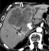
Hepatic Capsular Retraction. Axial contrast-enhanced CT demonstrates a large central mass forming CCA (block arrow) with hepatic capsular retraction (curved arrow) along the capsule of the liver. Also noted are dilated upstream bile ducts (thin arrow)
Fig. 12
Fig. 12
Lobulated Margins. Axial T1-weighted MRI in an early delayed phase demonstrates a peripheral mass forming CCA in the right hepatic lobe with lobulated margins (arrows) and rim enhancement
Fig. 13
Fig. 13
Necrosis. Axial contrast enhanced CT in the arterial phase (A), portal venous phase (B), and delayed phase (C), demonstrating a pathologically proven necrotic CCA with rim APHE (arrow), delayed central enhancement (curved arrow), and non-enhancing necrosis centrally (large arrow)

Similar articles

References

    1. Burnside ES, Sickles EA, Bassett LW, Rubin DL, Lee CH, Ikeda DM, et al. The ACR BI-RADS experience: learning from history. J Am Coll Radiol. 2009;6(12):851–60. doi: 10.1016/j.jacr.2009.07.023. PubMed PMID: 19945040; PubMed Central PMCID: PMC3099247. - PMC - PubMed
    1. Marks RM, Fung A, Cruite I, Blevins K, Lalani T, Horvat N, et al. The adoption of LI-RADS: a survey of non-academic radiologists. Abdom Radiol (NY). 2023;48(8):2514–24. Epub 20230526. doi: 10.1007/s00261-023-03951-9. PubMed PMID: 37233747. - PubMed
    1. Marks RM, Masch WR, Chernyak V. LI-RADS: Past, Present, and Future, From the AJR Special Series on Radiology Reporting and Data Systems. AJR Am J Roentgenol. 2021 Feb;216(2):295–304. 10.2214/AJR.20.24272. Epub 2020 Dec 23. PMID: 33052720. - PubMed
    1. Pesapane F, Tantrige P, De Marco P, Carriero S, Zugni F, Nicosia L, Bozzini AC, Rotili A, Latronico A, Abbate F, Origgi D, Santicchia S, Petralia G, Carrafiello G, Cassano E. Advancements in Standardizing Radiological Reports: A Comprehensive Review. Medicina (Kaunas). 2023 Sep 17;59(9):1679. 10.3390/medicina59091679. PMID: 37763797; PMCID: PMC10535385. - PMC - PubMed
    1. American College of Radiology Committee on LI-RADS®. The LI-RADS v2018 Manual 2018. Available from: https://www.acr.org/-/media/ACR/Files/Clinical-Resources/LIRADS/LI-RADS-.... Accessed on April 12, 2024.

MeSH terms

LinkOut - more resources