Clinical Trial
. 2025 Mar;31(3):861-868.
doi: 10.1038/s41591-024-03451-3. Epub 2025 Jan 7.

Intracerebroventricular B7-H3-targeting CAR T cells for diffuse intrinsic pontine glioma: a phase 1 trial

Affiliations
Clinical Trial

Intracerebroventricular B7-H3-targeting CAR T cells for diffuse intrinsic pontine glioma: a phase 1 trial

Nicholas A Vitanza et al. Nat Med. 2025 Mar.

Abstract

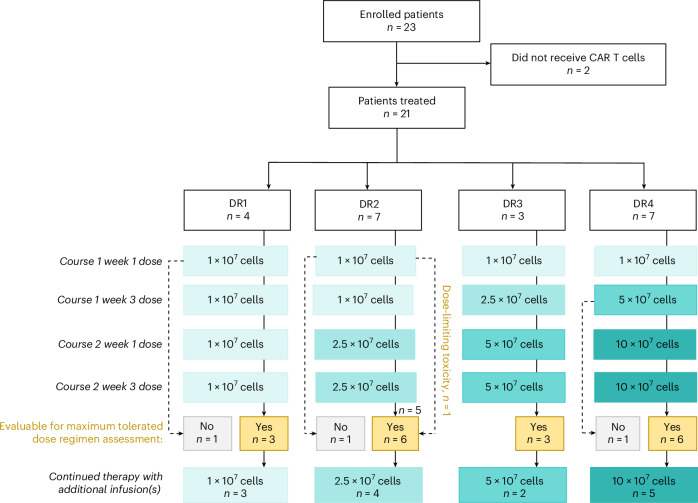
Diffuse intrinsic pontine glioma (DIPG) is a fatal central nervous system (CNS) tumor that confers a median survival of 11 months. As B7-H3 is expressed on pediatric CNS tumors, we conducted BrainChild-03, a single-center, dose-escalation phase 1 clinical trial of repetitive intracerebroventricular (ICV) dosing of B7-H3-targeting chimeric antigen receptor T cells (B7-H3 CAR T cells) for children with recurrent or refractory CNS tumors and DIPG. Here we report results from Arm C, restricted to patients with DIPG. The primary objectives were to assess feasibility and tolerability, which were both met. Secondary objectives included assessments of CAR T cell distribution and survival. A total of 23 patients with DIPG enrolled, and 21 were treated with repeated doses of ICV B7-H3 CAR T cells using intra-patient dose-escalation regimens without previous lymphodepletion. Concurrent tumor-directed therapy, including re-irradiation, was not allowed while on protocol therapy. We delivered a total of 253 ICV doses and established the highest planned dose regimen, DR4, which escalated up to 10 × 107 cells per dose, as the maximally tolerated dose regimen. Common adverse events included headache, fatigue and fever. There was one dose-limiting toxicity (intratumoral hemorrhage) during DR2. For all treated patients (n = 21), the median survival from their initial CAR T cell infusion was 10.7 months and the median survival from diagnosis was 19.8 months with 3 patients still alive at 44, 45 and 52 months from diagnosis. Ultimately, this completed first-in-human trial shows that repetitive ICV dosing of B7-H3 CAR T cells in pediatric and young adult patients with DIPG is tolerable, including multiyear repeated dosing, and may have clinical efficacy that warrants further investigation on a multisite phase 2 trial. ClinicalTrials.gov registration: NCT04185038 .


Conflict of interest statement

Competing interests: N.A.V. holds equity in and serves as the Scientific Advisory Board Chair for BrainChild Bio, Inc. J.A.G. holds equity in BrainChild Bio, Inc. R.J.O. receives research support from Lentigen Technology, a Miltenyi Biotec company, and is a consultant for Umoja Biopharma. R.A.G. is an inventor and receives royalties on patents related to CAR T cell technologies that are licensed to Juno Therapeutics, a Bristol Myers Squibb company, and serves as a consultant to Moonlight Bio. M.C.J. holds equity in and is the Chief Scientific Officer of BrainChild Bio, Inc. M.C.J. holds equity in, is a Board Observer for and serves as a member of the Joint Steering Committee of Umoja Biopharma, Inc. N.A.V., J.A.G., J.B.F., J.R.P. and M.C.J. are inventors on issued and pending patents related to CAR T cell therapies. The other authors declare no competing interests.

Figures

Fig. 1
Fig. 1. BrainChild-03 Arm C trial design.
CONSORT diagram of BrainChild-03 Arm C.
Fig. 2
Fig. 2. Survival following intracranial B7-H3 CAR T cells.
a, Swimmer plot describing patient history from time of diagnosis through death or most recent follow-up. For patients with multiple progressions or long-standing ongoing progression, only initial progression may be noted. b, Kaplan–Meier survival after the initial B7-H3 CAR T cell infusion for all treated patients. c, Kaplan–Meier survival stratified by previous progression status at the time of initial CAR T infusion. Shaded areas in b and c denote 95% confidence limits for the Kaplan–Meier estimates.
Fig. 3
Fig. 3. Neuroimaging after locoregional B7-H3 CAR T cell infusion.
a, Longitudinal MRIs of S021. T2-weighted (coronal top row, axial middle row) and axial post-contrast T1-weighted (bottom row) MRI images focused on the pontine lesion at various timepoints (see column labels). At diagnosis (DX), the pons is expanded with non-enhancing T2 hyperintensity, prepontine cistern effacement (dashed arrows) and partial effacement of the fourth ventricle (asterisks). Following radiation (Post RT, 4 months before immunotherapy (IT)), the lesion is smaller, with reduced prepontine cistern effacement (dashed arrows), persistent nodular T2 hyperintensity in the left dorsal pons (solid arrows) and biopsy- and therapy-related changes in the central pons (arrowheads). From the initiation of IT (Pre IT) to 30 months Post IT, the overall pons size has remained stable while hyperintensity has decreased. b, Longitudinal MRI of the pontine lesion of S053. Axial T2-weighted (top row) and axial post-contrast T1-weighted (bottom row) MRI images, focused on the pontine lesion at various timepoints (see column labels). At DX, the pons is enlarged (arrows) with diffuse T2 hyperintensity, heterogeneous enhancement and partial effacement of the fourth ventricle (asterisks). Following radiation (Post RT, 2 months before IT), there is further expansion of the pons and increased T2 signal abnormality (arrows), with persistent enhancement. From Pre IT to 2 months Post IT, the pontine lesion is smaller (arrows). However, by 4 months Post IT, the lesion is larger, with a new region of enhancement (arrowheads). At 6 and 8 months Post IT, the lesion size, T2 signal abnormality and enhancement are reduced.
Fig. 4
Fig. 4. Chemokine and cytokine concentrations in CSF during the DLT period.
a, A volcano plot of all 53 cytokines tested. The labels indicate cytokines showing at least a twofold change and a false discovery rate (FDR)-adjusted P < 0.05 across all four course–week combinations. b, A forest plot for six cytokines that show distinctive pre- and post-infusion patterns with subsequent infusions: those trending with cumulative infusions (CRP, SAA and TARC) and those consistently upregulated or downregulated after each infusion (IFNγ, CXCL10 (also known as IP-10) and GM-CSF). Data are presented as the model estimate of the post–pre-change on a log2 scale ± s.e.m. For differential analyses, only samples with matched pre- and post-infusion pairs at each Cr and W combination from the same patient were included. For the analysis presented in the volcano plot, the linear mixed model combined data from Cr1.W1 (n = 12), Cr1.W3 (n = 11), Cr2.W1 (n = 11) and Cr2.W3 (n = 10), resulting in 44 pre-infusion and 44 post-infusion measurements for each cytokine meeting quality filters. Study participants were included as random intercepts, and pre- and post-infusion status was included as a fixed effect. Analytes were measured in duplicate, samples with signal coefficients of variation greater than 25% were excluded and concentrations below the LLOD were considered undetectable (0 pg ml−1). Data were log2 transformed for analysis, and fold changes are presented on the log2 scale.
Extended Data Fig. 1
Pathologic diagnostic information for all patients.
Extended Data Fig. 2
Extended Data Fig. 2. Intracranial Detection of CAR T Cells in CSF.
Detection of B7-H3 CAR T cells in CSF pre- and post- infusion during the DLT period consists of 10 scheduled collections: Cr1.W1.Pre, Cr1.W1.Post, Cr1.W3.Pre, Cr1.W3.Post, Cr1.W4, Cr2.W1.Pre, Cr2.W1.Post, Cr2.W3.Pre, Cr2.W3.Post, Cr2.W4, which are defined as (where Cr: course; W: week; Pre: pre-infusion, and Post: post-infusion). The x-axis shows the sequence of these collections. Samples with lymphocytes count below the limit of quantitation (LOQ) were excluded from analysis. Shaded circles represent samples where B7-H3 CAR T cells were detected, defined by lymphocyte/EGFRt+% levels above the limit of detection (LOD) and a minimal count (>1) of detected EGFRt+ cells. The size of the circles denotes the percentage of EGFRt+ lymphocytes (Lymph/EGFRt+%) detected in the sample. Notably, S014 had no sample collected due to the placement of the shunt, S018 only received one dose, and S045 missed all post-infusion collection.
Extended Data Fig. 3
Extended Data Fig. 3. CSF CAR T cell detection Table.
Levels of CAR detection by EGFRt+ across all patients and across all dose regimens (DR1, DR2, DR3 and DR4) during the DLT period. Gating Strategy: Live> Singlets> Lymphocyte> CD3+> EGFRt+.
Extended Data Fig. 4
Extended Data Fig. 4. CSF cytokine data.
Absolute concentrations and pre-infusion versus post-infusion changes for cytokines exhibiting at least a two-fold change and a false discovery rate (FDR)-adjusted p-value < 0.05 in a linear mixed model. The model combined data from Cr1.W1 (n = 12), Cr1.W3 (n = 11), Cr2.W1 (n = 11), and Cr2.W3 (n = 10), resulting in n = 44 pre-infusion and n = 44 post-infusion measurements for each cytokine that met quality criteria. Subjects were included as random intercepts, and pre/post-infusion status was included as a fixed effect. Analytes were measured in duplicate; those with coefficients of variation (CV) greater than 25% were excluded. Concentrations were log2-transformed for analysis, and fold changes are presented on a linear scale. Subsequent columns show fold changes and adjusted p-values for the separate and combined infusion time points.
Extended Data Fig. 5
Extended Data Fig. 5. Representative flow gating strategy for CART cell detection.
Gating strategy used for flow-based CAR detection in CSF. Selection of the singlet, viable [CD36-] lymphocyte population was performed prior to T cell gating and exam­ination of the EGFRt+ CART cells and CD4/CD8 expression.

References

    1. Cooney, T. et al. Contemporary survival endpoints: an International Diffuse Intrinsic Pontine Glioma Registry study. Neuro. Oncol.19, 1279–1280 (2017). - PMC - PubMed
    1. Vitanza, N. A. & Monje, M. Diffuse intrinsic pontine glioma: from diagnosis to next-generation clinical trials. Curr. Treat. Options Neurol.21, 37 (2019). - PMC - PubMed
    1. Gardner, R. A. et al. Intent-to-treat leukemia remission by CD19 CAR T cells of defined formulation and dose in children and young adults. Blood129, 3322–3331 (2017). - PMC - PubMed
    1. Maude, S. L. et al. Chimeric antigen receptor T cells for sustained remissions in leukemia. N. Engl. J. Med.371, 1507–1517 (2014). - PMC - PubMed
    1. Vitanza, N. A. et al. Locoregional CAR T cells for children with CNS tumors: clinical procedure and catheter safety. Neoplasia36, 100870 (2023). - PMC - PubMed

Publication types

MeSH terms

Associated data