

Enhancing repeatability of follicle counting with deep learning reconstruction high-resolution MRI in PCOS patients
- PMID: 39775101
- PMCID: PMC11868616
- DOI: 10.1038/s41598-024-84812-3
Enhancing repeatability of follicle counting with deep learning reconstruction high-resolution MRI in PCOS patients
Abstract
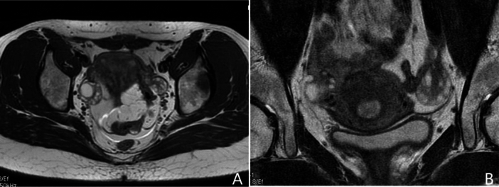
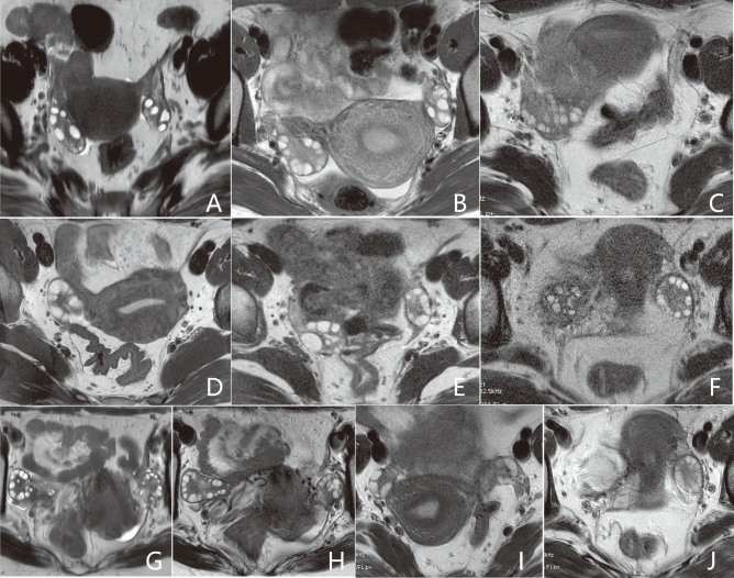
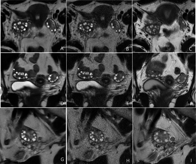
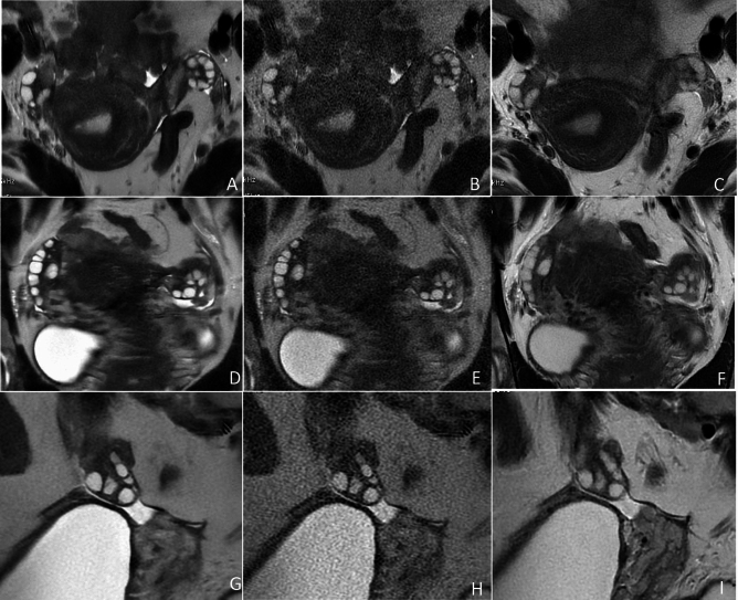
Follicle count, a pivotal metric in the adjunct diagnosis of polycystic ovary syndrome (PCOS), is often underestimated when assessed via transvaginal ultrasonography compared to MRI. Nevertheless, the repeatability of follicle counting using traditional MR images is still compromised by motion artifacts or inadequate spatial resolution. In this prospective study involving 22 PCOS patients, we employed periodically rotated overlapping parallel lines with enhanced reconstruction (PROPELLER) and single-shot fast spin-echo (SSFSE) T2-weighted sequences to suppress motion artifacts in high-resolution ovarian MRI. Additionally, deep learning (DL) reconstruction was utilized to compensate noise in SSFSE imaging. We compared the performance of DL reconstruction SSFSE (SSFSE-DL) images with conventional reconstruction SSFSE (SSFSE-C) and PROPELLER images in follicle detection, employing qualitative indices (blurring artifacts, subjective noise, and conspicuity of follicles) and the repeatability of follicle number per ovary (FNPO) assessment. Despite similar subjective noise between SSFSE-DL and PROPELLER as assessed by one observer, SSFSE-DL images outperformed SSFSE-C and PROPELLER images across all three qualitative indices, resulting in enhanced repeatability in FNPO assessment. These results highlighted the potential of DL reconstruction high-resolution SSFSE imaging as a more dependable method for identifying polycystic ovary, thus facilitating more accurate diagnosis of PCOS in future clinical practices.
Keywords: Deep learning; High resolution; Magnetic resonance imaging; Ovary; Polycystic ovary syndrome.
© 2025. The Author(s).
Conflict of interest statement
Declarations. Competing interests: The authors declare no competing interests.
Figures
Similar articles
-
High-Resolution Single-Shot Fast Spin-Echo MR Imaging with Deep Learning Reconstruction Algorithm Can Improve Repeatability and Reproducibility of Follicle Counting.J Clin Med. 2023 Apr 30;12(9):3234. doi: 10.3390/jcm12093234. J Clin Med. 2023. PMID: 37176674 Free PMC article.
-
Impact of Deep Learning Reconstruction Combined With a Sharpening Filter on Single-Shot Fast Spin-Echo T2-Weighted Magnetic Resonance Imaging of the Uterus.Invest Radiol. 2022 Jun 1;57(6):379-386. doi: 10.1097/RLI.0000000000000847. Epub 2022 Jan 10. Invest Radiol. 2022. PMID: 34999668
-
Effect of Deep Learning Reconstruction on Respiratory-triggered T2-weighted MR Imaging of the Liver: A Comparison between the Single-shot Fast Spin-echo and Fast Spin-echo Sequences.Magn Reson Med Sci. 2024 Apr 1;23(2):214-224. doi: 10.2463/mrms.mp.2022-0111. Epub 2023 Mar 29. Magn Reson Med Sci. 2024. PMID: 36990740 Free PMC article.
-
Definition and significance of polycystic ovarian morphology: a task force report from the Androgen Excess and Polycystic Ovary Syndrome Society.Hum Reprod Update. 2014 May-Jun;20(3):334-52. doi: 10.1093/humupd/dmt061. Epub 2013 Dec 16. Hum Reprod Update. 2014. PMID: 24345633
-
Ultrasonographic criteria in the diagnosis of polycystic ovary syndrome: a systematic review and diagnostic meta-analysis.Hum Reprod Update. 2024 Jan 3;30(1):109-130. doi: 10.1093/humupd/dmad027. Hum Reprod Update. 2024. PMID: 37804097 Free PMC article.
Cited by
-
Artificial intelligence in polycystic ovarian syndrome management: past, present, and future.Radiol Med. 2025 Jun 23. doi: 10.1007/s11547-025-02032-9. Online ahead of print. Radiol Med. 2025. PMID: 40549330 Review.
References
-
- Brown, M. A. & Chang, R. J. Polycystic ovary syndrome. Ultrasound Q.23, 233–238 (2007). - PubMed
-
- Walter, K. What is polycystic ovary syndrome?. JAMA327, 294 (2022). - PubMed
-
- Group T. R. E. A. Revised 2003 consensus on diagnostic criteria and long-term health risks related to polycystic ovary syndrome (PCOS). Hum. Reprod.19, 41–47 (2004). - PubMed
Publication types
MeSH terms
Grants and funding
LinkOut - more resources
Full Text Sources
Medical
