. 2025 Jan 7;41(1):23.
doi: 10.1007/s10565-024-09973-3.

Exosomal NEDD4L derived from HG+oxLDL-induced vascular endothelial cells accelerates macrophage M1 polarization and oxLDL uptake by ubiquitinating IκBα and PPARγ

Affiliations

Exosomal NEDD4L derived from HG+oxLDL-induced vascular endothelial cells accelerates macrophage M1 polarization and oxLDL uptake by ubiquitinating IκBα and PPARγ

Guozhu Chen et al. Cell Biol Toxicol. .

Abstract

Background: Vascular endothelial cell-derived exosomes are thought to mediate disease progression by regulating macrophage polarization. However, its mechanism in diabetes mellitus (DM)-related atherosclerosis (AS) progress is unclear.

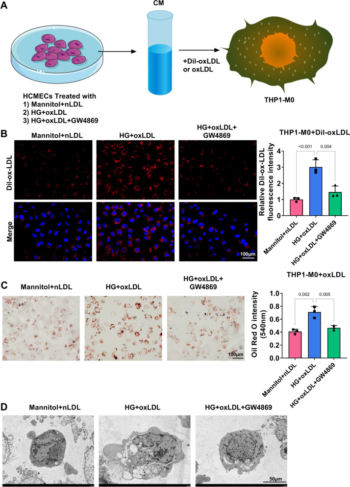
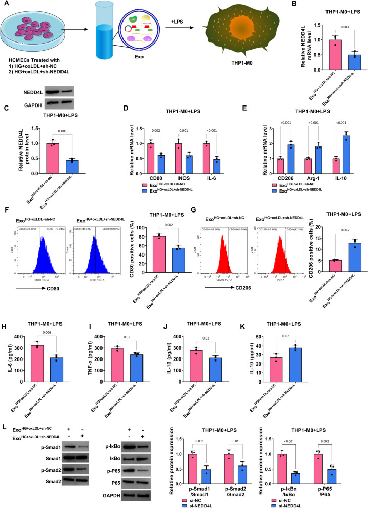
Methods: High-glucose (HG) and oxLDL were used to induce human cardiac microvascular endothelial cells (HCMECs) to mimic DM-related AS model. The conditioned medium (CM) from HG+oxLDL-induced HCMECs was incubated with THP1-M0 monocytes treated with LPS or oxLDL. The mRNA levels of macrophage M1/M2 polarization markers, NEDD4L, IκBα and PPARγ were determined by qRT-PCR. Flow cytometry was used to analyze macrophage marker. Dil-labeled oxLDL and oil red O staining were performed to assess oxLDL uptake by THP1-M0 cells. The levels of inflammatory factors were examined using ELISA. Transmission electron microscope was used for observing foam cell formation and exosome morphology. The protein levels of p-Smad1/Smad1, p-Smad2/Smad2, p-IκBα/IκBα, p-P65/P65, anti-lipid metabolism-related markers, and NEDD4L were tested by western blot. The interaction between NEDD4L and IκBα or PPARγ was assessed by Co-IP assay.

Results: The CM of HG+oxLDL-induced HCMECs could promote macrophage M1 polarization, oxLDL uptake and foam cell formation, and exosome inhibiter GW4869 eliminated these effects. NEDD4L was overexpressed in exosomes from HG+oxLDL-induced HCMECs, which could be taken up by THP1-M0 cells. Exosomal NEDD4L knockdown inhibited macrophage M1 polarization, oxLDL uptake and foam cell formation by reducing the protein levels of p-Smad1/Smad1, p-Smad2/Smad2, p-IκBα/IκBα and p-P65/P65. NEDD4L could reduce IκBα and PPARγ expression through ubiquitination.

Conclusion: HG+oxLDL-induced HCMECs-derived exosomal NEDD4L could enhance the ubiquitination of IκBα and PPARγ to facilitate macrophage M1 polarization and oxLDL uptake, thus accelerating DM-related AS.

Keywords: Atherosclerosis; Diabetes mellitus; Exosome; Macrophages; NEDD4L.


Conflict of interest statement

Declarations. Ethics approval and consent to participate: Not applicable. Conflicts of interest: The authors declare no competing interests.

Figures

Fig. 1
Fig. 1
Exosomes from HG+oxLDL-induced HCMECs mediated macrophage M1 polarization. A Flow chart of cell treatment and co-incubation. B-C qRT-PCR for detecting CD80, iNOS, IL-6, CD206, Arg-1 and IL-10 mRNA levels in LPS-induced THP1-M0 cells. D-E Flow cytometry for analyzing CD80 and CD206 positive cells in LPS-induced THP1-M0 cells. F-I ELISA was used to test the levels of IL-6, TNF-α, IL-1β and IL-10 in LPS-induced THP1-M0 cells. All experiments were performed in triplicate and P<0.05 was denoted statistically significant
Fig. 2
Fig. 2
Exosomes from HG+oxLDL-induced HCMECs mediated oxLDL uptake and foam cell formation. A Flow chart of cell co-incubation. B Fluorescence microscope was used to observe Dil-labeled oxLDL in THP1-M0 cells. C Oil red O staining to evaluate lipid deposition in oxLDL-induced THP1-M0 cells. D TEM for observing foam cell formation. All experiments were performed in triplicate and P<0.05 was denoted statistically significant
Fig. 3
Fig. 3
NEDD4L expression in exosomes from HG+oxLDL-induced HCMECs. A TEM for observing the microstructure of exosomes from HCMECs treated with Mannitol+nLDL or HG+oxLDL. B WB for testing TSG101, CD81, CD63 and Calnexin protein expression in exosomes from HCMECs treated with Mannitol+nLDL or HG+oxLDL. C NTA for detecting the particle sizes and concentrations of exosomes from HCMECs treated with Mannitol+nLDL or HG+oxLDL. D NEDD4L mRNA level was tested by qRT-PCR in HCMECs treated with Mannitol+nLDL or HG+oxLDL. E NEDD4L mRNA level in exosomes from HCMECs treated with Mannitol+nLDL or HG+oxLDL was measured by qRT-PCR. F Fluorescence microscope was used to observe Dil-labeled Exo in THP1-M0 cells after co-culturing with ExoMannitol+nLDL or ExoHG+oxLDL. G-H qRT-PCR and WB for testing NEDD4L mRNA and protein levels in THP1-M0 cells after co-culturing with ExoMannitol+nLDL or ExoHG+oxLDL. All experiments were performed in triplicate and P<0.05 was denoted statistically significant
Fig. 4
Fig. 4
Effect of exosomal NEDD4L knockdown from HG+oxLDL-induced HCMECs on macrophage M1 polarization. A Flow chart of cell treatment and co-incubation. B-C qRT-PCR and WB for detecting NEDD4L mRNA and protein levels in LPS-induced THP1-M0 cells. D-E qRT-PCR for testing CD80, iNOS, IL-6, CD206, Arg-1 and IL-10 mRNA levels in LPS-induced THP1-M0 cells. F-G CD80 and CD206 positive cells were analyzed by flow cytometry in LPS-induced THP1-M0 cells. H-K The levels of IL-6, TNF-α, IL-1β and IL-10 in LPS-induced THP1-M0 cells was analyzed by ELISA. L Protein levels of p-Smad1/Smad1, p-Smad2/Smad2, p-IκBα/IκBα and p-P65/P65 in LPS-induced THP1-M0 cells were examined by WB. All experiments were performed in triplicate and P<0.05 was denoted statistically significant
Fig. 5
Fig. 5
Effect of exosomal NEDD4L knockdown from HG+oxLDL-induced HCMECs on oxLDL uptake and foam cell formation. A Flow chart of cell co-incubation. B-C NEDD4L mRNA and protein levels in oxLDL-induced THP1-M0 cells were examined by qRT-PCR and WB. D Dil-labeled oxLDL in THP1-M0 cells was observed using fluorescence microscope. E Lipid deposition in oxLDL-induced THP1-M0 cells was assessed by Oil red O staining. F Foam cell formation was observed under TEM. G Protein levels of PPARγ, ABCG1, ABCA1 and LDLR in oxLDL-induced THP1-M0 cells were tested by WB. All experiments were performed in triplicate and P<0.05 was denoted statistically significant
Fig. 6
Fig. 6
Effect of NEDD4L on the ubiquitination of IκBα and PPARγ. A-B The mRNA and protein levels of NEDD4L, IκBα and PPARγ were detected by qRT-PCR and WB in THP1-M0 cells transfected with si-NC/si-NEDD4L. C-D WB was used for detected IκBα and PPARγ protein levels in THP1-M0 cells transfected with si-NC/si-NEDD4L and treated with CHX for indicated time points. E-F Co-IP for assessing the interaction between NEDD4L and IκBα or PPARγ. G-H Detection of ubiquitination levels of IκBα and PPARγ in THP1-M0 cells transfected with si-NC/si-NEDD4L. All experiments were performed in triplicate and P<0.05 was denoted statistically significant
Fig. 7
Fig. 7
The mechanism diagram of this study. Exosomal NEDD4L derived from HG+oxLDL-induced HCMECs could ubiquitinate IκBα/PPARγ and phosphorylate Smad1/Smad2, thus promoting macrophage M1 polarization, oxLDL uptake and foam cell formation

References

    1. Bai S, Yin Q, Dong T, Dai F, Qin Y, Ye L, et al. Endothelial progenitor cell-derived exosomes ameliorate endothelial dysfunction in a mouse model of diabetes. Biomed Pharmacother. 2020;131:110756. - PubMed
    1. Cai H, Pang Y, Wang Q, Qin W, Wei C, Li Y, et al. Proteomic profiling of circulating plasma exosomes reveals novel biomarkers of Alzheimer's disease. Alzheimers Res Ther. 2022;14(1):181. - PMC - PubMed
    1. Chen S, Li K, Guo J, Chen HN, Ming Y, Jin Y, et al. circNEIL3 inhibits tumor metastasis through recruiting the E3 ubiquitin ligase Nedd4L to degrade YBX1. Proc Natl Acad Sci USA. 2023;120(13):e2215132120. - PMC - PubMed
    1. Cheng P, Xie X, Hu L, Zhou W, Mi B, Xiong Y, et al. Hypoxia endothelial cells-derived exosomes facilitate diabetic wound healing through improving endothelial cell function and promoting M2 macrophages polarization. Bioact Mater. 2024;33:157–73. - PMC - PubMed
    1. Cui L, Ma J. NEDD4L Promotes IkappaBalpha Ubiquitination and Degradation in the Pathogenesis of Diabetic Retinopathy. Curr Eye Res. 2024;49(1):62–72. - PubMed

Publication types

MeSH terms

LinkOut - more resources