. 2025 Jan 7;36(1):5.
doi: 10.1007/s10856-024-06849-0.

Knockdown of decorin in human bone marrow mesenchymal stem cells suppresses proteoglycan layer formation and establishes a pro-inflammatory environment on titanium oxide surfaces

Affiliations

Knockdown of decorin in human bone marrow mesenchymal stem cells suppresses proteoglycan layer formation and establishes a pro-inflammatory environment on titanium oxide surfaces

Hisanobu Kamio et al. J Mater Sci Mater Med. .

Abstract

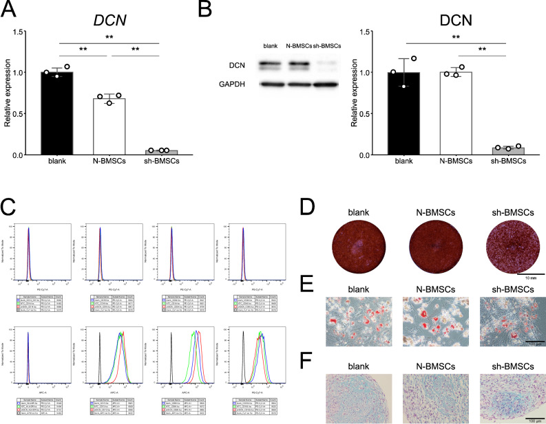
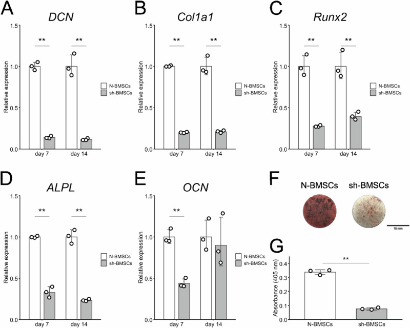
Osseointegration is essential for successful implant treatment. However, the underlying molecular mechanisms remain unclear. In this study, we focused on decorin (DCN), which was hypothesized to be present in the proteoglycan (PG) layer at the interface between bone and the titanium oxide (TiOx) surface. We utilized DCN RNA interference in human bone marrow mesenchymal stem cells (hBMSCs) to investigate its effects on PG layer formation, proliferation, initial adhesion, cell extension, osteogenic capacity, fibrotic markers, and immunotolerance to TiOx in vitro. After 14 days of cultivation, we observed no PG layer was detected, and the osteogenic capacity was suppressed in DCN-depleted hBMSCs. Furthermore, the conditioned medium upregulated the expression of M1 macrophage markers in human macrophages. These results suggest that endogenous DCN plays a crucial role in PG layer formation and that the PG layer alters inflammation around Ti materials.


Conflict of interest statement

Compliance with ethical standards. Conflict of interest: The authors declare no competing interests.

Figures

Fig. 1
Fig. 1
Characterization of N-BMSCs and sh-BMSCs. A mRNA and B protein expression analysis of DCN. C Representative flow cytometry analysis of cell surface markers. D Alizarin red S staining, E Oil Red O staining, and F Alcian blue staining after osteogenic induction, adipogenic induction, and chondrogenic induction culture. **p < 0.01
Fig. 2
Fig. 2
A Transmission electron micrograph of the cell–TiOx interface. The ruthenium-positive areas were detected at 14 days of N-BMSC cultivation (arrowheads). B Proliferation of N-BMSCs and sh-BMSCs on TiOx on days 1, 2, 3, 7, and 14. C Initial cell adhesion of N-BMSCs and sh-BMSCs to TiOx at 1, 2, and 3 h. This figure is representative of three independent experiments. **p < 0.01
Fig. 3
Fig. 3
Immunofluorescence staining analysis of N-BMSCs and sh-BMSCs after 24 h of cultivation on TiOx. A Expression of vinculin. Green: F-actin, Red: vinculin, Blue: DAPI. B Area, C major, D minor, E aspect ratio, F Feret, and G minimum Feret were measured to evaluate cell adhesion and extension. **p < 0.01
Fig. 4
Fig. 4
Osteogenic capacity of N-BMSCs and sh-BMSCs. Gene expression of A DCN, B Col1a1, C Runx2, D ALPL, and E OCN after 7 and 14 days of cultivation on TiOx. Mineralized nodule formation by alizarin red S staining (F) and quantitative analysis (G). This figure is representative of three independent experiments. **p < 0.01
Fig. 5
Fig. 5
Gene expression of A Acta2, B FN1, and C TGF-β1 after 7 and 14 days of cultivation on TiOx. Western blotting analysis of extracellular matrix after 14 days of cultivation on TiOx (D) and semi-quantification (EH). This figure is representative of two independent experiments. *p < 0.05, **p < 0.01
Fig. 6
Fig. 6
Gene expression related to immunotolerance in N-BMSCs and sh-BMSCs after 14 days of TiOx (AC) cultivation and in CM-treated hMps derived from N-BMSCs and sh-BMSCs (DJ). Protein expression levels of IL-6 and IL-10 in the supernatant of BMSCs cultured on TiOx (K, L) and after stimulating hMps (M, N). **p < 0.01
Fig. 7
Fig. 7
Decellularization assay analysis of N-BMSCs and sh-BMSCs. Gene expression of A Col1a1, B Runx2, C ALPL, D OCN, E Acta2, F FN1, and G TGF-β1 after 14 days of cultivation on decellularized TiOx. SEM-EDX analysis of decellularized Ti surfaces (H, I). This figure is representative of two independent experiments. *p < 0.05, **p < 0.01

Similar articles

References

    1. Albrektsson T, Johansson C. Osteoinduction, osteoconduction and osseointegration. Eur Spine J. 2001;10(Suppl 2):S96–101. 10.1007/s005860100282 - PMC - PubMed
    1. Huang J, Zhang X, Yan W, Chen Z, Shuai X, Wang A, et al. Nanotubular topography enhances the bioactivity of titanium implants. Nanomedicine. 2017;13:1913–23. 10.1016/j.nano.2017.03.017 - PubMed
    1. Kopf BS, Schipanski A, Rottmar M, Berner S, Maniura-Weber K. Enhanced differentiation of human osteoblasts on Ti surfaces pre-treated with human whole blood. Acta Biomater. 2015;19:180–90. 10.1016/j.actbio.2015.03.022 - PubMed
    1. Sverzut AT, Crippa GE, Morra M, de Oliveira PT, Beloti MM, Rosa AL. Effects of type I collagen coating on titanium osseointegration: histomorphometric, cellular and molecular analyses. Biomed Mater. 2012;7:035007. 10.1088/1748-6041/7/3/035007 - PubMed
    1. Jiang J, Wan F, Yang J, Hao W, Wang Y, Yao J, et al. Enhancement of osseointegration of polyethylene terephthalate artificial ligament by coating of silk fibroin and depositing of hydroxyapatite. Int J Nanomedicine. 2014;9:4569–80. 10.2147/IJN.S69137 - PMC - PubMed

MeSH terms

LinkOut - more resources