

Monitoring the impact of confinement on hyphal penetration and fungal behavior
- PMID: 39775212
- PMCID: PMC11684722
- DOI: 10.1371/journal.pone.0312855
Monitoring the impact of confinement on hyphal penetration and fungal behavior
Abstract
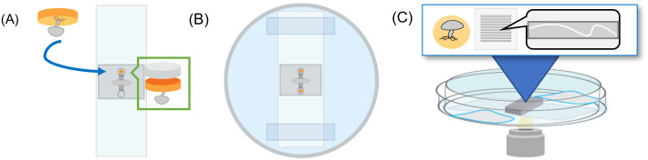
Through their expansive mycelium network, soil fungi alter the physical arrangement and chemical composition of their local environment. This can significantly impact bacterial distribution and nutrient transport and can play a dramatic role in shaping the rhizosphere around a developing plant. However, direct observation and quantitation of such behaviors is extremely difficult due to the opacity and complex porosity of the soil microenvironment. In this study, we demonstrate the development and use of an engineered microhabitat to visualize fungal growth in response to varied levels of confinement. Microfluidics were fabricated using photolithography and conventional soft lithography, assembled onto glass slides, and prepared to accommodate fungal cultures. Selected fungal strains across three phyla (Ascomycota: Morchella sextalata, Fusarium falciforme; Mucoromycota: Linnemannia elongata, Podila minutissima, Benniella; Basidiomycota: Laccaria bicolor, and Serendipita sp.) were cultured within microhabitats and imaged using time-lapse microscopy to visualize development at the mycelial level. Fungal hyphae of each strain were imaged as they penetrated through microchannels with well-defined pore dimensions. The hyphal penetration rates through the microchannels were quantified via image analysis. Other behaviors, including differences in the degree of branching, peer movement, and tip strength were also recorded for each strain. Our results provide a repeatable and easy-to-use approach for culturing fungi within a microfluidics platform and for visualizing the impact of confinement on hyphal growth and other fungal behaviors pertinent to their remodeling of the underground environment.
Copyright: © 2024 Guo et al. This is an open access article distributed under the terms of the Creative Commons Attribution License, which permits unrestricted use, distribution, and reproduction in any medium, provided the original author and source are credited.
Conflict of interest statement
The authors have declared that no competing interests exist.
Figures


References
-
- Howe A, Bonito G, Chou MY, Cregger MA, Fedders A, Field JL, et al.. Frontiers and Opportunities in Bioenergy Crop Microbiome Research Networks. Vol. 6, Phytobiomes Journal. American Phytopathological Society; 2022. p. 118–26.
-
- Vetterlein D, Carminati A, Kögel-Knabner I, Bienert GP, Smalla K, Oburger E, et al.. Rhizosphere Spatiotemporal Organization—A Key to Rhizosphere Functions. Frontiers in Agronomy. 2020;2(July):1–22.
-
- Nunan N. Game Changer in Soil Science The microbial habitat in soil: Scale, heterogeneity and functional. 2017;425–9.
MeSH terms
LinkOut - more resources
Full Text Sources
