. 2025 Jan 7;20(1):e0315673.
doi: 10.1371/journal.pone.0315673. eCollection 2025.

Revealing the impact of tadalafil-loaded proniosomal gel against dexamethasone-delayed wound healing via modulating oxido-inflammatory response and TGF-β/Macrophage activation pathway in rabbit model

Affiliations

Revealing the impact of tadalafil-loaded proniosomal gel against dexamethasone-delayed wound healing via modulating oxido-inflammatory response and TGF-β/Macrophage activation pathway in rabbit model

Nermin A Helmy et al. PLoS One. .

Abstract

A serious challenge of the chronic administration of dexamethasone (DEX) is a delay in wound healing. Thus, this study aimed to investigate the potential of Tadalafil (TAD)-loaded proniosomal gel to accelerate the healing process of skin wounds in DEX-challenged rabbits. Skin wounds were induced in 48 rabbits of 4 groups (n = 12 per group) and skin wounds were treated by sterile saline (control), TAD-loaded proniosomal gel topically on skin wound, DEX-injected rabbits, and DEX+TAD-loaded proniosomal gel for 4 weeks. The optical photography, transmission electron microscopy, in vitro release profile, and stability studies revealed the successful preparation of the selected formula with good stability. DEX administration was associated with uncontrolled oxido-inflammatory reactions, suppression in immune response in skin wounds, and consequently failure in the healing process. TAD-loaded proniosomal gel-treated rabbits manifested a marked enhancement in the rate of wound closure than control and DEX groups (p < 0.05). The TAD-loaded proniosomal gel successfully antagonized the impacts of DEX by dampening MDA production, and enhancing total antioxidant capacity, coupled with modulation of inflammatory-related genes, inducible nitric oxide synthase, tumor necrosis factor-alpha, interleukin-1β, and matrix metalloproteinase 9, during all healing stages (p < 0.05). This was in combination with significant amplification of immune response-related genes, CD68 and CD163 (p < 0.05). Moreover, the histopathological, Masson's Trichrome-stain, and immune-histochemical studies indicated a successful tissue recovery with the formation of new blood vessels in groups treated with TAD-loaded proniosomal gel, as manifested by well-organized collagen fibers, upregulation of transforming growth factor β1, and vascular endothelial growth factor immune expression in skin tissues (p < 0.05). Overall, the topical application of TAD-loaded proniosomal gel is useful in improving the delayed wound healing linked to DEX therapy via regulating the release of inflammatory/macrophage activation mediators and enhanced antioxidant capacity, angiogenesis, and vascularity.


Conflict of interest statement

The authors have declared that no competing interests exist.

Figures
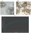
Fig 1
Fig 1. Optical photography (A), Transmission electron microscopy (B), and release profile of the chosen formula (C).
Fig 2
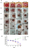
Fig 2. (A) Gross examination of wound area of the experimental groups, and (B) Wound diameter during the experiment period for the experimental groups.
Data are presented as mean ± SD.
Fig 3
Fig 3. Changes in mRNA expression of iNOS (A) TNF-α (B) IL-1β (C), and MMP-9 (D) genes in the different groups. Data are expressed as mean ± SE (n = 4).
Means with distinct letters in the same column are significantly dissimilar at p < 0.05.
Fig 4
Fig 4. Changes in expression of macrophage activation-related genes, CD68 (A) and CD163 (B) in the experimental groups.
Data are expressed as mean ± SE (n = 4). Means with distinct letters in the same column are significantly dissimilar at p<0.05.
Fig 5
Fig 5
Photomicrograph of skin wound (H&E-stained sections), (a) control group on day 7 showed intense inflammatory cell infiltration and necrosis, (b) TAD group on day 7 showed intense inflammatory cell infiltration with necrosis, (c) DEX group, on day 7 showed intense inflammatory cell infiltration, edema, and scab formation, (d) TAD+DEX group at day 7 showing scab covered wound with intense inflammatory cell infiltration, (e) The control group on day 14 showed scab-covered wounds consisting of necrotic tissue and inflammatory cells, underlying tissue showing inflammatory cell infiltration, and unorganized granulation tissue, (f) TAD group on day 14 showed fewer inflammatory cell infiltration with well-arranged organized tissue, (g) DEX group, on day 14 showed intense inflammatory cell infiltration with necrosis, (h) TAD+DEX group on day 14 showed few inflammatory cells and organized tissue formation, (i) The control group on day 21 showed moderate inflammatory cell infiltration, and unorganized granulation tissue with no evidence of re-epithelialization, (j) The TAD group on day 21 showed re-epithelialization with scarce inflammatory cell infiltration and well-formed granulation tissue, (k) DEX group, on day 21 showed moderate inflammatory cell infiltration with unorganized granulation tissue, and (l) TAD+DEX group at day 21 showing mild inflammatory cell infiltration, and re-epithelialization with granulation tissue formation. (scale bar 100 μm).
Fig 6
Fig 6
Photomicrograph of skin wound (MTC stained sections), (a) control group at day 7 showed few collagen fibers in the wound area. (b) The TAD group on day 7 showed a moderate amount of collagen. (c) DEX group, on day 7 showed few collagen fibers. (d) the TAD+DEX group on day 7 showed few collagen fibers formation. (e) The control group on day 14 showed few collagen fibers in the wound area. (f) The TAD group on day 14 showed an increase in the amount of collagen fiber. (g) DEX group, on day 14 showed disorganized collagen fibers. (h) TAD+DEX group on day 14 showed increased collagen formation. (i) The control group on day 21 showed moderate disoriented collagen fiber. (j) The TAD group on day 21 showed a high amount of well-organized collagen fiber. (k) DEX group, on day 21 showing disorganized collagen fibers. (l) The TAD+DEX group on day 21 showed moderate collagen fiber amount. (scale bar 100 μm). (m) area % of collagen fiber in different treated groups (data was expressed as mean ± SE, different letters indicating significant differences at p < 0.05). TAD; Tadalafil-Loaded Proniosomal Gel, DEX; Dexamethasone.
Fig 7
Fig 7
Photomicrograph of skin wound (TGF-β1 stained sections), (a) control group at day 7 showing weak immune expression. (b) The TAD group on day 7 showed moderate immune expression. (c) DEX group, on day 7 showed weak immune expression. (d) TAD+DEX group on day 7 showed weak immune expression. (e) The control group on day 14 showed moderate immune expression. (f) The TAD group on day 14 showed moderate immune expression. (g) DEX group, on day 14 showed moderate immune expression. (h) TAD+DEX group on day 14 showed moderate immune expression. (i) Control group at day 21 showing moderate immune expression (j) TAD group at day 21 showing strong immune expression. (k) DEX group, on day 21 showed moderate immune expression. (l) the TAD+DEX group on day 21 showed strong immune expression. (scale bar 100 μm). (m) area % of TGF-β1 expression in different treated groups (data was expressed as mean ± SE, different letters indicating significant differences at p < 0.05). TAD; Tadalafil-Loaded Proniosomal Gel, DEX; Dexamethasone.
Fig 8
Fig 8. Photomicrograph of skin wound (VEGF-stained sections).
(a) the control group on day 7 showed weak immune expression. (b) The TAD group at day 7 showed moderate immune expression. (c) DEX group, on day 7 showed weak immune expression. (d) TAD+DEX group on day 7 showed weak immune expression. (e) The control group on day 14 showed moderate immune expression. (f) The TAD group on day 14 showed moderate immune expression. (g) DEX group, on day 14 showed moderate immune expression. (h) TAD+DEX group on day 14 showed moderate immune expression. (i) the control group on day 21 showed moderate immune expression (j) the TAD group on day 21 showed strong immune expression. (k) DEX group, on day 21 showing moderate immune expression. (l) TAD+DEX group on day 21 showing strong immune expression. (scale bar 100 μm). (m) area % of VEGF expression in different treated groups (data was expressed as mean ± SE, different letters indicating significant differences at p < 0.05). TAD; Tadalafil-Loaded Proniosomal Gel, DEX; Dexamethasone.

Similar articles

Cited by

References

    1. Armstrong DG, Meyr AJ, Sanfey H, Eidt JF, Mills JL, Billings JA. Basic principles of wound management. Atlas Small Anim Wound Manag Reconstr Surgery; John Wiley Sons; Hoboken, NJ, USA. 2018; 33–52.
    1. Borena BM, Martens A, Broeckx SY, Meyer E, Chiers K, Duchateau L, et al.. Regenerative skin wound healing in mammals: state-of-the-art on growth factor and stem cell based treatments. Cell Physiol Biochem. 2015;36: 1–23. doi: 10.1159/000374049 - DOI - PubMed
    1. Rajendran NK, Kumar SSD, Houreld NN, Abrahamse H. A review on nanoparticle based treatment for wound healing. J Drug Deliv Sci Technol. 2018;44: 421–430.
    1. Reinke JM, Sorg H. Wound repair and regeneration. Eur Surg Res. 2012;49: 35–43. doi: 10.1159/000339613 - DOI - PubMed
    1. Zhao R, Liang H, Clarke E, Jackson C, Xue M. Inflammation in chronic wounds. Int J Mol Sci. 2016;17: 2085. doi: 10.3390/ijms17122085 - DOI - PMC - PubMed

MeSH terms

LinkOut - more resources