. 2025 Jan 7;16(1):17.
doi: 10.1007/s12672-025-01741-x.

Histologically confirmed pediatric extracardiac rhabdomyoma: case series

Affiliations

Histologically confirmed pediatric extracardiac rhabdomyoma: case series

Gashaw Arega et al. Discov Oncol. .

Abstract

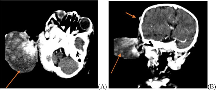
Rhabdomyoma is a rare benign tumor of striated muscle, which can be either cardiac or extracardiac. Extracardiac rhabdomyomas can occur throughout the body, though the fetal and adult subtypes are most commonly found in the head and neck region.We present three pediatric cases of extracardiac rhabdomyoma, fetal subtype, detailing their clinical presentations, computed tomography imaging, and tissue biopsy findings. Given the very rare occurrence of extracardiac rhabdomyoma and its relatively benign nature, histological diagnosis is crucial. In all three cases reported here, a diagnosis of extracardiac rhabdomyoma was confirmed, and treatment with local excision resulted in favorable outcomes.

Keywords: Pediatrics; Retroperitoneum; Rhabdomyoma; Surgery.


Conflict of interest statement

Declarations. Ethics approval and consent to participate: The study was approved by the Addis Ababa University, Department’s of Pediatrics Research and Ethics Committee and Institutional Review Board, and the study was conducted in accordance with the WMA Declaration of Helsinki. Consent for publication: Written informed consent was obtained from the patient’s parents for anonymized patient information to be published in this article. Competing interests: The authors declare no competing interests.

Figures

Fig. 1
Fig. 1
post-contrast neck Ct images sagittal A coronal B and axial C, All the selected images demonstrate soft tissue mass that has homogenous attenuation at retropharyngeal space (orange arrow) with no significant calcification, necrosis, or adjacent bone destruction
Fig. 2
Fig. 2
A grey white well-circumscribed mass. B: Well-circumscribed periphery (40×magnification). C: Fascicular arrangement of strap cells, (arrows) 400×magnification
Fig. 3
Fig. 3
Post-contrast axial A and coronal B brain CT, both images demonstrate well-demarcated soft tissue mass located at the mastoid area that has increased central flow with no bone destruction or extension to intracranial space (orange arrow). A small scalp lesion is noted that has no imaging appearance of aggressive (B -small arrow)
Fig. 4
Fig. 4
10 × objective microscopy showing spindle cells with ovoid nuclei interspersed with rhabdoid cells. B: 40 × objective, Hematoxylin and Eosin section, large neoplastic cells with pinkish fibrillary cytoplasm and small nuclei. C: Immunohistochemistry: Muscle-specific actin positive in the large polygonal cells. D: Immunohistochemistry showed Desmin diffuse expression
Fig. 4
Fig. 4
10 × objective microscopy showing spindle cells with ovoid nuclei interspersed with rhabdoid cells. B: 40 × objective, Hematoxylin and Eosin section, large neoplastic cells with pinkish fibrillary cytoplasm and small nuclei. C: Immunohistochemistry: Muscle-specific actin positive in the large polygonal cells. D: Immunohistochemistry showed Desmin diffuse expression
Fig. 5
Fig. 5
AD: Upper retroperitoneal well-defined mass (arrow) A-pre-contrast image and B-post-contrast image showing homogenous contrast enhancement. C-axial and D- sagittal image showing a well-defined homogenously enhancing midline upper retroperitoneal mass measuring 2.3 cm × 2.3 cm × 1.8 cm in size

References

    1. Zolota V, Tzelepi V, Charoulis N, et al. Mediastinal rhabdomyoma: case report and review of the literature. Virchows Arch. 2006;449:124–8. 10.1007/s00428-006-0211-8. - PubMed
    1. Sciacca P, Giacchi V, Mattia C, et al. Rhabdomyomas and tuberous sclerosis complex: our experience in 33 cases. BMC Cardiovasc Disord. 2014;14:66. 10.1186/1471-2261-14-66. - PMC - PubMed
    1. Kapadia SB, Meis JM, Frisman DM, Ellis GL, Heffner DK, Hyams VJ. Adult rhabdomyoma of the head and neck: a clinicopathologic and immunophenotypic study. Hum Pathol. 1993. 10.1016/0046-8177(93)90240-H. - PubMed
    1. Willis J, Abdul-Karim FW, di Sant’Agnese PA. Extracardiac rhabdomyomas. Semin Diagn Pathol. 1994;11(1):15–25. - PubMed
    1. Sagar N, Mandal S, Khurana N, Kumar A. Cervical rhabdomyoma a rare entity: case report with review of literature. Indian J Pathol Microbiol. 2020;63(3):467–9. 10.4103/IJPM.IJPM_73_19. - PubMed

LinkOut - more resources