. 2025 Jan 7;21(1):e1012804.
doi: 10.1371/journal.ppat.1012804. eCollection 2025 Jan.

SARS-CoV-2-induced cytokine storm drives prolonged testicular injury and functional impairment in mice that are mitigated by dexamethasone

Affiliations

SARS-CoV-2-induced cytokine storm drives prolonged testicular injury and functional impairment in mice that are mitigated by dexamethasone

Stefanos Giannakopoulos et al. PLoS Pathog. .

Abstract

Compromised male reproductive health, including reduced testosterone and sperm count, is one of the long COVID symptoms in individuals recovering from mild-severe disease. COVID-19 patients display testicular injury in the acute stage and altered serum fertility markers in the recovery phase, however, long-term implications on the testis remain unknown. This study characterized the consequences of SARS-CoV-2 on testis function. The K18-hACE2 mice that survived SARS-CoV-2 infection were followed for one month after infection and the testicular injury and function markers were assessed at different stages of infection and recovery. The long-term impact of infection on key testes function-related hormones and male fertility was measured. The efficacy of inflammation-suppressing drug in preventing testicular injury was also evaluated. The morphological defects like sloughing of spermatids into the lumen and increased apoptotic cells sustained for 2-4 weeks after infection and correlated with testicular inflammation and immune cell infiltration. Transcriptomic analysis revealed dysregulation of inflammatory, cell death, and steroidogenic pathways. Furthermore, reduced testosterone levels associated with a transient reduction in sperm count and male fertility. Most testicular impairments resolved within one month of infection. Importantly, dexamethasone treatment attenuated testicular damage, inflammation, and immune infiltration. Our results implicate virus-induced cytokine storm as the major driver of testicular injury and functional impairments, timely prevention of which limits testis damage. These findings serve as a model for evaluating therapeutics in long COVID patients and may guide clinical strategies to improve male reproductive health outcomes post-SARS-CoV-2 infection.


Conflict of interest statement

The authors have declared that no competing interests exist.

Figures

Fig 1
Fig 1. Testis morphological defects in SARS-CoV-2-infected K18-hACE2 mice.
The days post-infection (D) are represented as pre-symptomatic (D3), symptomatic (D5), short-term recovery (D8-14), and long-term recovery stages (D30). (A) Representative images of PAS-H-stained testis sections from mock (Ctrl) and infected mice. Leydig cells, white arrows; Sertoli cells, black arrows. Inset shows normal round and elongating spermatids. Various testicular abnormalities were observed, including the occurrence of apoptotic germ cells (white arrowheads in D8), apoptotic meiotic cells (black arrowheads in D8), cells with degenerating nuclei (short white arrow in D8), tubules lacking clear lumen and with a separation of germ cell layer from the basal membrane (D5), and with the sloughing of healthy spermatids and spermatocytes into the lumen (D8-14). The stages of seminiferous epithelium are: II-IV (D30), V-VI (Ctrl & D3) VII-VIII (D5 & D8), X-XII (D14). Inset, 3x magnification. Scale, 100 μm. (B) Quantification of seminiferous tubule organizational and germ cell defects. Each data point corresponds to a different mouse. For each male, 50 tubules were examined. Statistical significance (t-test, Ctrl vs. Infected). *p<0.05 and **p<0.01. The differences approaching significance (p = 0.05–0.1) are shown directly in the graph.
Fig 2
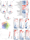
Fig 2. RNA-seq analysis of the testis identifies dysregulation of distinct transcriptional pathways at different stages of SARS-CoV-2 infection.
(A) Volcano plots of differentially expressed genes (DEGs) (n, 3 mice per time point) at indicated time points, dotted lines represent cutoff: p< 0.05 and log2fc > |1|, genes increased after infection, red; and genes decreased after infection, blue. (B) Venn diagram of total DEGs at D3 (red), D5 (yellow), D8 (light green), D14 (dark green), and D30 (blue). (C) Principal component analysis (PCA) performed using DEGs (p <0.05 and log2fc > |1|) and normalized transcripts per million (controls, black circle; infected, red circle) (D) Heatmap of top pathways and select diseases and functions revealed by Ingenuity Pathway Analysis (IPA), filtered to remove erroneous results (upregulated, red; downregulated, blue). (E) The expression log2 ratio of upregulated (red) and downregulated (blue) genes associated with ILK, FAK, and activin/inhibin signaling pathways and pathogen-induced cytokine storm, fibrosis, and apoptosis signaling pathways as determined by IPA in each dataset (p< 0.05; log2fc > |1|) at indicated time points.
Fig 3
Fig 3. Sustained increase in the levels of pro-inflammatory cytokine and chemokine in the testis correlate with systemic inflammation.
(A) Heatmap of a panel of COVID-19 specific cytokine and chemokines measured using LUMINEX assay in the tissue homogenates (lung, testis, and heart) and serum of K18-hACE2 mice at indicated time points post-infection (n, 4 mice per group). Data were normalized to the control, log10 transformed, and expressed as relative abundance (red, increased; blue, decreased; and white represents unchanged compared to uninfected controls). (B) Levels of IL-1β, IL-6, CXCL1, and TNF-α expressed as pg/mL. Individual data points represent different mice. The statistical significance (*p<0.05, **p<0.01, ***< p<0.001) was determined using Mann-Whitney test for all analyses (C) Pearson’s correlation coefficient analysis between testicular defects (cells in lumen, lack of lumen, and lamina separation) and key cytokines (red box) and chemokines (yellow box) in the serum (left) and testis (right) at D3, D5, and D8. Pearson coefficient >0.6 was considered a significant correlation.
Fig 4
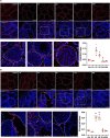
Fig 4. SARS-CoV-2 infection results in sustained apoptotic cell death in the testis.
Representative images of (A) TUNEL staining and (B) ZO-1 staining in testis from the indicated time point. Nuclei were visualized using DAPI stain (blue), and dotted lines represent the shape of individual seminiferous tubules. ImageJ analysis of (C) TUNEL staining normalized to respective DAPI positive cells and (D) ZO-1staining intensity. Each data point is the average of counts from 3 different fields from each testis section (n, 3 mice/time point). Significance (*p<0.05, **p<0.01, ***p<0.001) was determined using a student’s t-test.
Fig 5
Fig 5. SARS-CoV-2 infection leads to a transient increase in the infiltration of CD68+ and CD11b+ cells in the testis.
Representative images of testis sections at indicated days after infection (D). The DAPI (blue) and infiltrating leukocytes were visualized using (A) CD68, and (B) CD11b markers. Dotted lines outline the shape of individual seminiferous tubules and ImageJ analysis of CD68 and CD11b staining normalized to DAPI-positive cells was done using particle analyzer tool. Each data point is the average of counts from 3 different fields from each testis section (n, 3 mice/time point). Images were taken using Leica SP3, DMI8 confocal microscope. Significance (*p<0.05, **p<0.01, ***p<0.001) was determined using the student’s t-test.
Fig 6
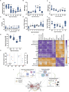
Fig 6. Consequences of SARS-CoV-2 infection on testis function.
Serum levels of (A) testosterone, (B) follicle-stimulating hormone (FSH), (C) inhibin B (INHB), and (D) luteinizing hormone (LH) were measured using ELISA at indicated time points and expressed as ng/mL serum (n, at least 4 mice per group). TGF-β levels in (E) serum and (F) testis homogenates were measured using ELISA and expressed as ng/mL at indicated time points post-infection. (G) Sperm count per epididymis was measured at indicated time points (n, 3–5 mice per group). Each data point represents data from one epididymis. (H) Age-matched male survivors were mated with females (2 females per male) for 6 days and then separated. The males were sacrificed after 6 days to measure the sperm count while pregnancy was evaluated after 10 days by counting the embryos. The percent change in the sperm count/epididymis (left y-axis, circle, blue) in each male as compared to un-infected controls and the number of fetuses (right y-axis, triangle, red) in each female (n, 2 females for each male). (I) Pearson’s correlation coefficient analysis of testicular defects, pro-inflammatory and cell death markers, and fertility parameters during the acute and short-term recovery stages (D3, D5, and D8). Significance (*p<0.05, **p<0.01, ***p<0.001, ****p<0.0001) was determined using student’s t-test. (J) Schematic illustration of the proposed events leading to testicular injury generated using BioRender.com. SARS-CoV-2 infection increases serum levels of pro-inflammatory cytokines/chemokines that trigger testicular inflammation leading to downstream effects including loss of BTB integrity, infiltration of immune cells, and apoptosis. These events manifest in the disruption of the morphology of the seminiferous tubules and impaired testicular function marked by hormone dysregulation and lower sperm count. Most of these impairments resolve within two spermatogenesis cycles, however, depending on the severity of these impairments, infection may lead to a short-term decrease in fertility in mice recovering from acute infection.
Fig 7
Fig 7. Effect of dexamethasone treatment on the testicular injury in SARS-CoV-2 infected mice.
Infected mice were treated with 10mg/kg dexamethasone starting D2 for 3 days and at D5 (A) SARS-CoV-2 genome copies in the lung measured using qRT-PCR and expressed as copies/mg RNA. The mRNA fold change of (B) IL6 and TNFA gene expression in the testis from infected mice with and without dexamethasone treatment using qRT-PCR at D5 (C) IL-6 and TNF-α levels in the lungs, serum, and testis measured using ELISA at D5 (D) Representative images of PAS-H-stained testis sections from mock (Ctrl), infected (D5), and dexamethasone-treated (Dex-D5) mice. Various testicular abnormalities were observed including the occurrence of apoptotic germ cells (black arrowheads) and apoptotic meiotic cells (white arrowheads), lack of lumen, separation from the basal membrane (*), and interstitial edema (IE). In Dex-D5 testes few minor defects were observed, exemplified here as 2 apoptotic meiotic cells (white arrowheads). Scale, 100 μm. For quantification of seminiferous tubule organizational and germ cell defects, 50 tubules were examined in each male. Each data point corresponds to a different mouse. (E) Representative images of the testis from SARS-CoV-2-infected mice at D5 with or without dexamethasone treatment with nuclei stained using DAPI (blue) and infiltrating leukocytes stained using antibodies against CD68 (top panel; red), and anti-CD11b (bottom panel; red). Dotted lines outline the shape of individual seminiferous tubules. (F) Representative images of the testis from mice with or without dexamethasone treatment with nuclei stained using DAPI (blue) and apoptotic cell visualized using TUNEL (red). ImageJ analysis of (G) TUNEL and (H) CD68 and CD11b staining normalized to DAPI staining measured using particle analyzer tool. Each data point represents the mean of three fields from different testis sections (n, 4 mice per group). Images were taken using Leica SP3, DMI8 confocal microscope. (I) Sperm count per epididymis was measured at D5 in the presence or absence of dexamethasone treatment and PBS control (n, 4–5 mice per group). Each data point represents data from one epididymis. Significance (*p<0.05, **p<0.01, ***p<0.001, ****p<0.0001) was determined using student’s t-test.

References

    1. Al-Aly Z, Xie Y, Bowe B. High-dimensional characterization of post-acute sequelae of COVID-19. Nature. 2021;594: 259–264. doi: 10.1038/s41586-021-03553-9 - DOI - PubMed
    1. Chen C, Haupert SR, Zimmermann L, Shi X, Fritsche LG, Mukherjee B. Global Prevalence of Post-Coronavirus Disease 2019 (COVID-19) Condition or Long COVID: A Meta-Analysis and Systematic Review. J Infect Dis. 2022;226: 1593–1607. doi: 10.1093/infdis/jiac136 - DOI - PMC - PubMed
    1. Lopez-Leon S, Wegman-Ostrosky T, Ayuzo Del Valle NC, Perelman C, Sepulveda R, Rebolledo PA, et al.. Long-COVID in children and adolescents: a systematic review and meta-analyses. Sci Rep. 2022;12: 9950. doi: 10.1038/s41598-022-13495-5 - DOI - PMC - PubMed
    1. Davis HE, McCorkell L, Vogel JM, Topol EJ. Long COVID: major findings, mechanisms and recommendations. Nat Rev Microbiol. 2023;21: 133–146. doi: 10.1038/s41579-022-00846-2 - DOI - PMC - PubMed
    1. Subramanian A, Nirantharakumar K, Hughes S, Myles P, Williams T, Gokhale KM, et al.. Symptoms and risk factors for long COVID in non-hospitalized adults. Nat Med. 2022;28: 1706–1714. doi: 10.1038/s41591-022-01909-w - DOI - PMC - PubMed

LinkOut - more resources