. 2025 Jan 8;20(1):e0311037.
doi: 10.1371/journal.pone.0311037. eCollection 2025.

In vitro and in vivo evaluation of Ulva lactuca for wound healing

Affiliations

In vitro and in vivo evaluation of Ulva lactuca for wound healing

Chien-Hsing Wang et al. PLoS One. .

Abstract

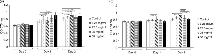
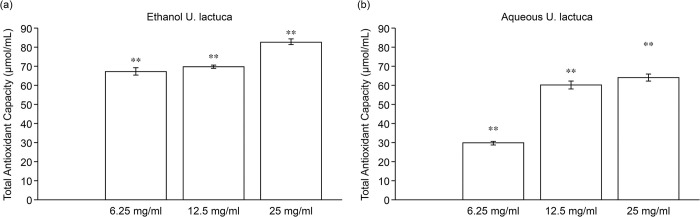
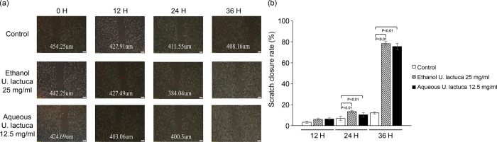
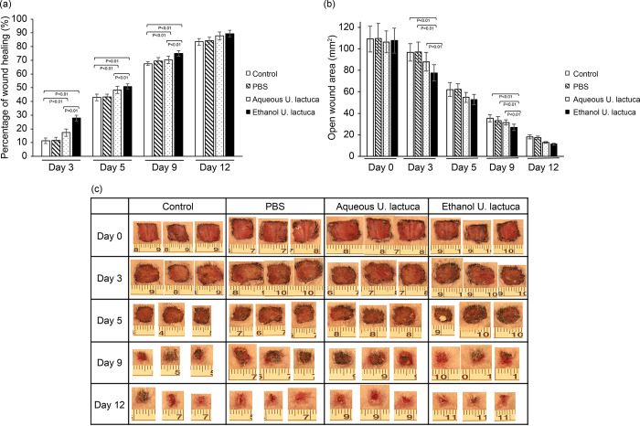
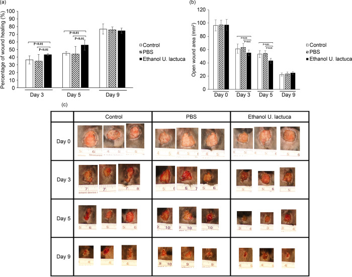
Ulva lactuca (U. lactuca) is an important seaweed species. Some ingredients in this species are thought to accelerate wound healing. However, limited data on the use of seaweed for wound healing exists. This study examined whether ethanol or aqueous extracts of U. lactuca promote antioxidant and anti-inflammatory properties in vitro and wound healing in vitro and in vivo. Cell proliferation, antioxidation, and migration were observed in NIH3T3 cells treated with U. lactuca extract in vitro. Both U. lactuca extracts were examined for their ability to inhibit inflammatory cytokine synthesis in lipopolysaccharide (LPS)-stimulated RAW 264.7 cells. In vivo experiments involved four groups of albino mice (BALB/c; 10 mice per group). One 1.0 cm2 wound was created via excision of full-thickness skin on the back of all mice. Group I mice were treated topically with the ethanol extract of U. lactuca (25 mg/mL) for 10 d. Group II mice were treated topically with an aqueous extract of U. lactuca (12.5 mg/mL) for 10 d. Group III mice received topical application of phosphate-buffered saline solution. Group IV mice wounds were maintained without treatment. Both extracts considerably increased fibroblast proliferation. The antioxidant activity of the U. lactuca extract was determined using a total antioxidant capacity assay. Both extracts inhibited the release of tumor necrosis factor-α (TNF-α) and interferon-γ (IFN-γ) from LPS-mediated inflammation in RAW 264.7 cells. These extracts also upregulated the expression of Th2 cytokines such as transforming growth factor beta 1 (TGF-β1) and interleukin 10 (IL-10) in RAW 264.7 cells under pro-inflammatory conditions. Both extracts enhanced the migratory ability of NIH3T3 cells. U. lactuca ethanol extract enhances wound healing properties in vivo. These results suggest that bioactive compounds derived from U. lactuca extract are beneficial for wound healing and anti-inflammatory therapies.


Conflict of interest statement

The authors have declared that no competing interests exist.

Figures

Fig 1
Fig 1. Ulva lactuca increases the proliferation rate of fibroblasts.
One set of 96-well culture dishes was seeded with NIH3T3 cells. Cells were treated with serial concentrations of ethanol (A) or aqueous (B) U. lactuca extract for 24 h. At 24 h intervals, cell numbers were assayed using the MTT method. Cells were analyzed in triplicate for each sample. *p <0.01 vs. control. All experiments were performed in triplicates, and the results consistently showed the same trend. Data presented represent one of three independent experiments.
Fig 2
Fig 2. Ulva. lactuca extract promotes antioxidant ability.
Total antioxidant capacity results for the U. lactuca extract using the CheKine™ Total Antioxidant Capacity (TAC) Assay Kit. Concentrations of 6.25, 12.5, and 25 mg/mL of the ethanol (A) or aqueous (B) extracts were evaluated for antioxidant activity. Absorbance was measured at 593 nm using a microplate reader. All experiments were performed in triplicates (n = 3). Values represent the mean ± S.D. Asterisks indicate statistical significance based on Student’s t tests (**P <0.01).
Fig 3
Fig 3. Ulva lactuca inhibits mRNA expression of pro-inflammatory cytokines in RAW 264.7.
Quantitative analysis of mRNA levels of pro-inflammatory cytokines. The values from treated RAW 264.7 cells were normalized to match GAPDH measurements and subsequently expressed as a ratio to mRNA in the control cells (n = 4 per group). *p <0.01 vs. control.
Fig 4
Fig 4. Ulva lactuca increases NIH3T3 migration.
Representative image of NIH3T3 cell migration. In vitro wound healing assay. The scratched NIH3T3 cells seeded on 6-well plates were cultured in DMEM in the presence of 25 mg/mL U. lactuca ethanol extract or 12.5 mg/mL U. lactuca aqueous extract. The number of cells in the scratched areas increased differently among the four groups at 0, 12, 24, and 36 h. (A) The photographs are representative of three independent experiments. (B) A qualitative scratch assay was performed. The scratch closure rate (%) was obtained by averaging the results of three independent experiments. The bars represent the mean of three independent experiments. P <0.05 indicates that averaging differed significantly between two different groups (paired Student’s t test). Scale bar represents 200 μm.
Fig 5
Fig 5. Ulva lactuca extract treatment promotes wound contraction in BALB/c mice.
A 1.0 cm2 cutaneous excisional lesion was created on the back skin of mice. Group I mice were treated with 25 mg/mL of U. lactuca ethanol extract three times daily. Excisional lesions of group II mice were treated with 12.5 mg/mL U. lactuca aqueous extract three times daily. Group III received PBS treatment, and group IV included control mice with untreated wounds. The size of the cutaneous lesions was measured using calipers on days 0, 3, 5, 9, and 12. Each group consisted of 10 mice. (A) Changes in the wound area at each time point relative to the original wound area of mice in each group over 12 d. The bars represent the percentage of wound healing in each group. p <0.05 indicates that the percentage of wound healing differed significantly between the two groups (paired Student’s t-test). Percentage of wound healing = (total wound area − present wound area)/(total wound area) × 100. (B) Changes in wound area at each time point in each group over 12 d. (C) Digital photographs of mice at various stages of wound healing. The photographs show representative results of the wounds on days 0, 3, 5, 9, and 12 after wound creation. Photographs represent three out of ten mice.
Fig 6
Fig 6. The ethanol extract of Ulva lactuca promotes wound contraction of C57BL/6 mice.
A 1.0 cm2 cutaneous excisional wound was created on the backs of mice. Group I mice were treated with 25 mg/mL of the U. lactuca ethanol extract three times daily. Group II mice were treated with PBS following excisional lesions. Group three comprised control mice with untreated wounds. Cutaneous wound size was measured using calipers on days 0, 3, 5, and 9. Each group consisted of 10 mice. (A) Changes in the wound area at each time point relative to the original wound area of mice in each group over 9 d. The bars represent the percentage of wound healing in each group. p <0.05 indicates that the percentage of wound healing differed significantly between the two groups (paired Student’s t-test). Percentage of wound healing = (Total wound area − Present wound area)/(Total wound area) × 100. (B) Changes in wound area at each time point of mice in each group over 9 d. (C) Digital photographs of mice at various stages of wound healing. The photographs show representative results of the wounds on days 0, 3, 5, and 9 after wound creation. Photographs represent three out of ten mice.

Similar articles

References

    1. Walker M. Human skin through the ages. Int J Pharm. 2022;622: 121850. doi: 10.1016/j.ijpharm.2022.121850 - DOI - PubMed
    1. Abazari M, Ghaffari A, Rashidzadeh H, Badeleh SM, Maleki Y. A systematic review on classification, identification, and healing process of burn wound healing. Int J Low Extrem Wounds. 2022;21: 18–30. doi: 10.1177/1534734620924857 - DOI - PubMed
    1. Haalboom M. Chronic wounds: Innovations in diagnostics and therapeutics. Curr Med Chem. 2018;25: 5772–5781. doi: 10.2174/0929867324666170710120556 - DOI - PubMed
    1. Shen S, Chen X, Shen Z, Chen H. Marine polysaccharides for wound dressings application: An overview. Pharmaceutics. 2021;13. doi: 10.3390/pharmaceutics13101666 - DOI - PMC - PubMed
    1. Wang JP, Ruan JL, Cai YL, Luo Q, Xu HX, Wu YX. In vitro and in vivo evaluation of the wound healing properties of Siegesbeckia pubescens. J Ethnopharmacol. 2011;134: 1033–1038. doi: 10.1016/j.jep.2011.02.010 - DOI - PubMed

Supplementary concepts

LinkOut - more resources