. 2024 Dec 10;9(2):ziae160.
doi: 10.1093/jbmrpl/ziae160. eCollection 2025 Feb.

Discoidin Domain Receptor 1 impacts bone microarchitecture with aging in female mice

Affiliations

Discoidin Domain Receptor 1 impacts bone microarchitecture with aging in female mice

Kimberly Denman et al. JBMR Plus. .

Abstract

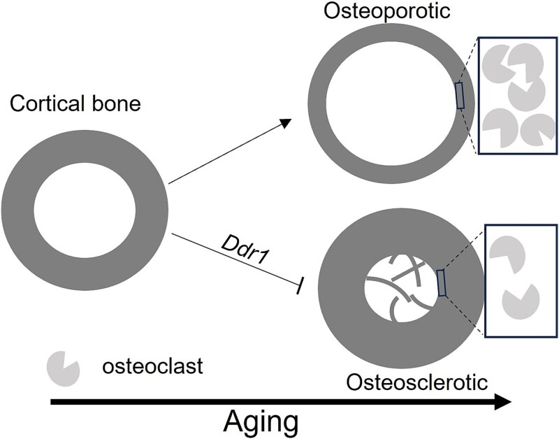
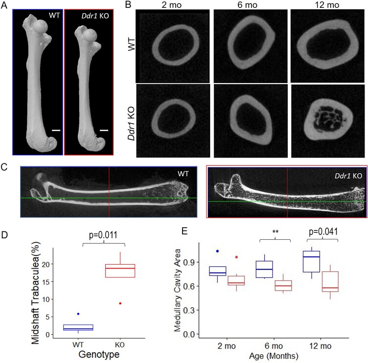
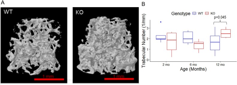
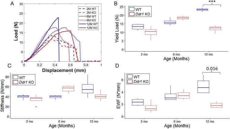
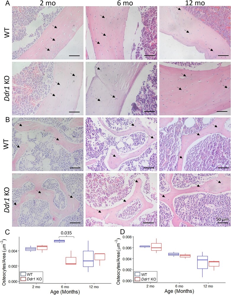
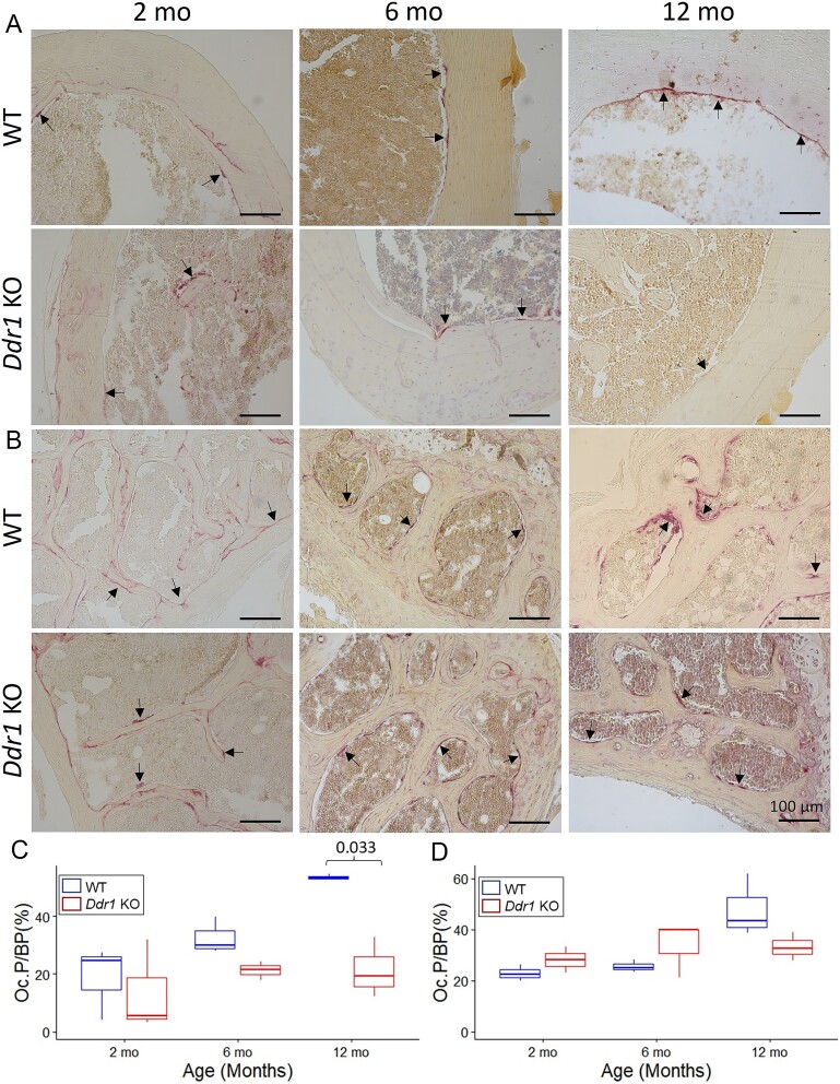
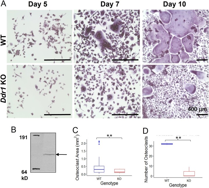
Discoidin Domain Receptor 1 (DDR1) is a receptor tyrosine kinase that binds to and is activated by collagen(s), including collagen type I. Ddr1 deletion in osteoblasts and chondrocytes has previously demonstrated the importance of this receptor in bone development. In this study, we examined the effect of DDR1 ablation on bone architecture and mechanics as a function of aging. Femurs were collected from female global Ddr1 knockout (KO) and wild-type (WT) mice at 2, 6, and 12 mo of age and analyzed by high-resolution micro-computed tomography (μCT), mechanical testing, and histology. Primary monocytes were collected for in vitro osteoclastogenesis assays. Our studies on younger (2 mo) mice revealed no significant differences between the two genotypes and the microarchitectural and mechanical features had a similar trend as those reported earlier for osteoblast or chondrocyte specific Ddr1 knockdown. At an advanced age (12 mo), significant differences were noted across the two genotypes. μCT analysis showed a decrease in medullary cavity area as well as increased trabeculation in cortical and trabecular bone in the Ddr1 KO vs. WT mice. In addition, Ddr1 KO mouse bones exhibited reduced mechanical properties (lower peak load, yield load, and energy to yield) at 12 mo. Histological analysis revealed reduced osteoclast count in Ddr1 KO femurs at 12 mo with no significant difference in osteocyte count between the genotypes. In vitro, osteoclastogenesis was impaired in Ddr1 KO bone marrow derived cells. These results suggest that DDR1 deficiency adversely impacts osteoclast differentiation and bone remodeling in an age-dependent manner.

Keywords: aging; biomechanics; bone microct; genetic animal model; osteoclasts.


Conflict of interest statement

None declared.

Figures

Graphical Abstract
Graphical Abstract
Figure 1
Figure 1
μCT of cortical bone. Representative whole femurs from Ddr1 KO and WT mice at 2 mo (A). Trabeculae growth was observed in the medullary cavity of 12-mo KO mice, as shown in cross-sections of the mid-femoral diaphysis (B) and longitudinal sections (C) of the murine femur. Quantitative analysis revealed a significant increase of trabecular growth in the mid-shaft cross-sections in 12-mo KO mice (D). The mid-femoral diaphysis also showed a decrease in medullary cavity area in KO femurs at 6 and 12 mo (**p<.01) (E). (Number of mice (n) for each group are listed in Table 1). Abbreviations: KO, knockout; μCT, micro-computed tomography; WO, wild-type.
Figure 2
Figure 2
μCT of trabecular bone. 3D reconstruction of trabecular regions of the femur at 12 mo (A) revealed increased trabecular number in 12-mo-old Ddr1 KO femurs as compared to WT (B). (Number of mice (n) for each group are listed in Table 1). Abbreviations: KO, knockout; μCT, micro-computed tomography; WO, wild-type.
Figure 3
Figure 3
Three-point bending mechanics. Representative three-point bending curves from 2-, 6- and 12-mo-old WT and Ddr1 KO femurs (A). Yield load and stiffness ascertained from three-point bending curves show an age-dependent increase for WT but a sharp decline for KO femurs at 12 mo of age (B, C). The energy to yield load (elastic work to failure) was calculated via the area under the curve and showed a similar trend (D). (***<.001) Number of mice used for each group was n = 4. Abbreviations: KO, knockout; WO, wild-type.
Figure 4
Figure 4
Osteocyte count. Representative H&E images for (A) cortical and (B) trabecular bone were used to ascertain osteocyte count. The osteocyte count per unit area was only found to be significantly higher in the cortical bone of 6-mo-old WT mice (C) and not in the trabecular bone (D). Number of mice used for each group was n = 3. Abbreviations: H&E, hematoxylin and eosin; WO, wild-type.
Figure 5
Figure 5
Osteoclast count. TRAP staining of cortical (A) and trabecular bone (B) revealed significantly decreased Oc.P. per BP in the cortical bone (C) at 12 mo but not in the trabecular bone (D). Number of mice used for each group was n = 3. Abbreviations: BP, bone perimeter; Oc.P., osteoclast perimeter; TRAP, tartrate-resistant acid phosphatase.
Figure 6
Figure 6
Osteoclastogenesis in vitro. TRAP staining of primary bone marrow cells from 6-mo-old female mice undergoing osteoclastogenesis in vitro (A). Western blot of whole cell lysates from osteoclasts differentiated from WT cells shows Ddr1 expression as a band of ~120 kD (B). Quantitative analysis showed that at day 10, multinucleated giant osteoclasts derived from KO cells were smaller in size (C) and fewer in number (D) as compared to those from WT cells (**p<.01). These in-vitro experiments were repeated for cells derived from at least n = 3 mice per genotype. Abbreviations: KO, knockout; TRAP, tartrate-resistant acid phosphatase; WT, wild-type.

Similar articles

References

    1. Dempster D, Felsenberg D, Geest S, eds. The Bone Quality Book: A Guide to Factors Influencing Bone Strength. Excerpta Medica Elsevier; 2006.
    1. Henriksen K, Leeming DJ, Byrjalsen I, et al. . Osteoclasts prefer aged bone. Osteoporos Int. 2007;18(6):751–759. 10.1007/s00198-006-0298-4 - DOI - PubMed
    1. Chung P-L, Zhou S, Eslami B, Shen L, LeBoff MS, Glowacki J. Effect of age on regulation of human osteoclast differentiation. J Cell Biochem. 2014;115(8):1412–1419. 10.1002/jcb.24792 - DOI - PMC - PubMed
    1. Cui J, Shibata Y, Zhu T, Zhou J, Zhang J. Osteocytes in bone aging: advances, challenges, and future perspectives. Ageing Res Rev. 2022;77:101608. 10.1016/j.arr.2022.101608 - DOI - PubMed
    1. Agarwal G, Smith AW, Jones B. Discoidin Domain Receptors: micro insights into macro assemblies. Biochim Biophys acta Mol cell Res. 2019;1866(11):118496. 10.1016/j.bbamcr.2019.06.010 - DOI - PMC - PubMed