

The effect of prenatal fumonisin B exposure on bone innervation in newborn Wistar rats
- PMID: 39776677
- PMCID: PMC11702245
- DOI: 10.2478/jvetres-2024-0056
The effect of prenatal fumonisin B exposure on bone innervation in newborn Wistar rats
Abstract
Introduction: This study explored the effects of prenatal exposure to fumonisins B (FB) on bone innervation in newborn Wistar rats.
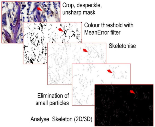
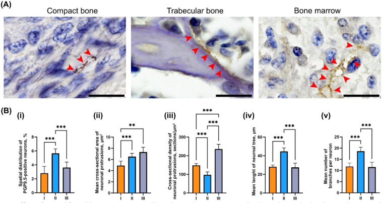
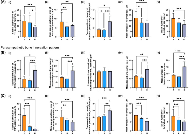
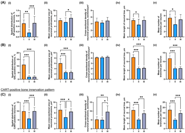
Material and methods: Pregnant dams (n = 6 per group) were assigned to either the control or one of two FB-exposed groups (60 mg or 90 mg/kg body weight) from the 7th day of gestation until parturition. On the day of parturition, one male pup from each litter (n = 6 per group) was randomly selected and euthanised, and their femurs were dissected for analysis. Bone innervation was quantified by examining the morphology patterns of sympathetic, parasympathetic, sensory and cocaine- and amphetamine-regulated transcript (CART)-positive fibres. Prepared bone sections were analysed using immunohistochemistry staining for protein gene product 9.5, tyrosine hydroxylase, choline acetyltransferase, vasoactive intestinal peptide (VIP), substance P and CART-positive neurons.
Results: The group that received a higher dose of FB demonstrated an increase in both the size and complexity of the complete bone neuronal network together with heightened sympathetic and sensory innervation, and displayed a decrease in neuron density and sympathetic innervation. Fumonisin B exposure led to a decrease in galanin-positive and VIP-positive bone neuronal networks in both groups exposed to FB, while in the lower-dose group, there was also a decrease in CART-positive innervation.
Conclusion: Prenatal FB exposure significantly influences the neuronal bone network of rats, which is essential for maintaining bone homeostasis. These findings emphasise the necessity for further research to understand the lasting effects and underlying mechanisms of alterations induced by FB.
Keywords: bone innervation; mycotoxins; peripheral nervous system; prenatal exposure.
© 2024 Ewa Tomaszewska et al., published by Sciendo.
Conflict of interest statement
Conflict of Interests Statement: The authors declare that there is no conflict of interests regarding the publication of this article.
Figures
Similar articles
-
Alterations of neurochemical expression of the coeliac-superior mesenteric ganglion complex (CSMG) neurons supplying the prepyloric region of the porcine stomach following partial stomach resection.J Chem Neuroanat. 2016 Mar;72:25-33. doi: 10.1016/j.jchemneu.2015.12.011. Epub 2015 Dec 28. J Chem Neuroanat. 2016. PMID: 26730724
-
The Influence of Prolonged Acetylsalicylic Acid Supplementation-Induced Gastritis on the Neurochemistry of the Sympathetic Neurons Supplying Prepyloric Region of the Porcine Stomach.PLoS One. 2015 Nov 25;10(11):e0143661. doi: 10.1371/journal.pone.0143661. eCollection 2015. PLoS One. 2015. PMID: 26606050 Free PMC article.
-
Nonselective innervation of lamina I projection neurons by cocaine- and amphetamine-regulated transcript peptide (CART)-immunoreactive fibres in the rat spinal dorsal horn.Eur J Neurosci. 2009 Jun;29(12):2375-87. doi: 10.1111/j.1460-9568.2009.06773.x. Epub 2009 May 22. Eur J Neurosci. 2009. PMID: 19490082
-
NTP technical report on the toxicity studies of Dibutyl Phthalate (CAS No. 84-74-2) Administered in Feed to F344/N Rats and B6C3F1 Mice.Toxic Rep Ser. 1995 Apr;30:1-G5. Toxic Rep Ser. 1995. PMID: 12209194
-
Histomorphometrical changes in intestine structure and innervation following experimental fumonisins intoxication in male Wistar rats.Pol J Vet Sci. 2020 Mar;23(1):77-88. doi: 10.24425/pjvs.2020.132751. Pol J Vet Sci. 2020. PMID: 32233304
References
-
- Association of Official Analytical Chemists AOAC Method 2001.04-2001, Fumonisins B1 and B2 in corn and corn flakes. 19th. AOAC International; Gaithersburg, MD, USA: 2012. : , edition, , , .
-
- Collins T.F.X., Sprando R.L., Black T.N., Shackelford M.E., Laborde J.B., Hansen D.K., Eppley R.M., Trucksess M.W., Howard P.C., Bryant M.A., Ruggles D.I., Olejnik N., Rorie J.I. Effects of Fumonisin B1 in Pregnant Rats Part 2. Food Chem Toxicol. 1998;36:673. doi: 10.1016/S0278-6915(98)00036-2. :, . , , –. , doi: . - DOI - PubMed
-
- Devreese M., De Backer P., Croubels S. Overview of the most important mycotoxins for the pig and poultry husbandry Vlaam Diergeneeskd Tijdschr. 2013;82:171. doi: 10.21825/vdt.v82i4.16694. :, . , , –. , doi: . - DOI
LinkOut - more resources
Full Text Sources