

Double Outlet Right Ventricle: A Rare Finding in a 15-Month-Old Female With Failure to Thrive
- PMID: 39776777
- PMCID: PMC11705528
- DOI: 10.1002/ccr3.70076
Double Outlet Right Ventricle: A Rare Finding in a 15-Month-Old Female With Failure to Thrive
Abstract
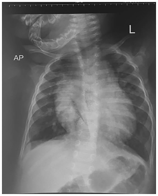
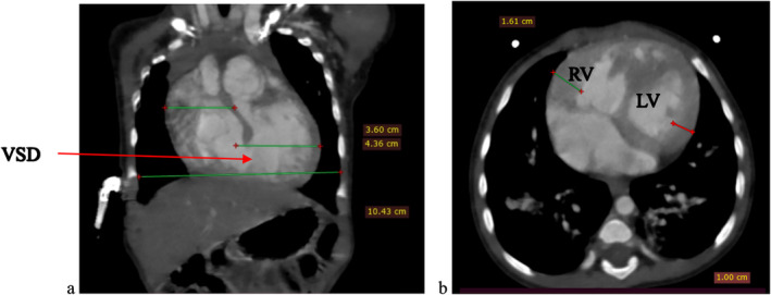
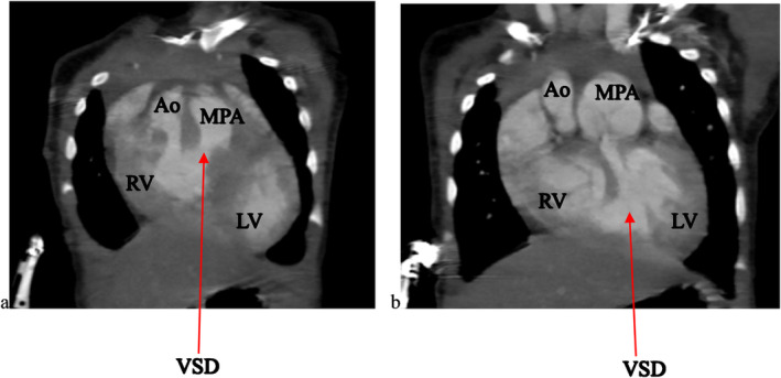
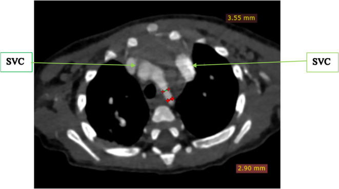
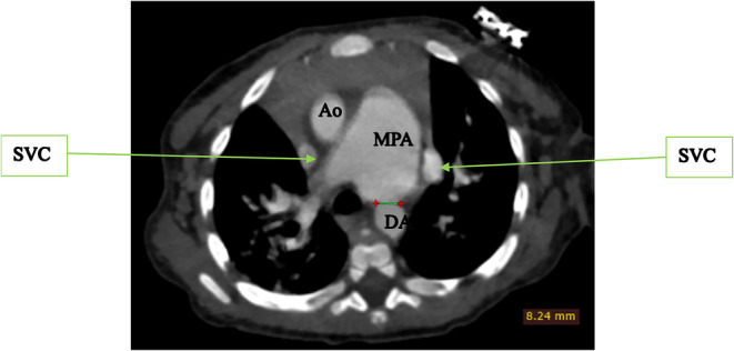
Double outlet right ventricle (DORV) is a rare congenital heart defect where both the aorta and pulmonary artery originate from the right ventricle, often accompanied by additional cardiac anomalies to mitigate circulatory imbalance, though such compensations usually fail. We report a 15-month-old infant with recurrent respiratory infections and poor weight gain, referred for computed tomography angiography. Physical examination showed a small, non-syndromic infant with pallor, tachypnea, irritability, and finger clubbing. Initial imaging revealed cardiomegaly and lung infiltrates; echocardiography and computed tomography angiography confirmed additional intracardiac defects of double superior vena cavae, a hypoplastic aortic arch, hypertrophic right ventricular wall, and a patent ductus arteriosus. Corrective surgery was delayed due to respiratory complications. This case emphasizes the critical need to consider cardiac pathology in pediatric patients with recurrent respiratory symptoms, as untreated DORV can lead to high mortality.
Keywords: cardiac computed tomography; case report; congenital heart defect; double outlet right ventricle; echocardiography.
© 2025 The Author(s). Clinical Case Reports published by John Wiley & Sons Ltd.
Conflict of interest statement
The authors declare no conflicts of interest.
Figures
References
-
- Karev E. and Stovpyuk O. F., “Double Outlet Right Ventricle in Adults: Anatomic Variability, Surgical Treatment, and Late Postoperative Complications,” Journal of Clinical Ultrasound 50, no. 8 (2022): 1151–1165. - PubMed
-
- Obler D., Juraszek A. L., Smoot L. B., and Natowicz M. R., “Double Outlet Right Ventricle: Aetiologies and Associations,” Journal of Medical Genetics 45, no. 8 (2008): 481–497. - PubMed
-
- Farooqi K. M., Uppu S. C., Nguyen K., et al., “Application of Virtual Three‐Dimensional Models for Simultaneous Visualization of Intracardiac Anatomic Relationships in Double Outlet Right Ventricle,” Pediatric Cardiology 37, no. 1 (2016): 90–98. - PubMed
LinkOut - more resources
Full Text Sources
