

Management of an Intraparenchymal Struvite Stone Using Laparoscopic Nephrolithotomy: A Case Report
- PMID: 39776779
- PMCID: PMC11705428
- DOI: 10.1002/ccr3.70065
Management of an Intraparenchymal Struvite Stone Using Laparoscopic Nephrolithotomy: A Case Report
Abstract
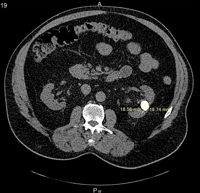
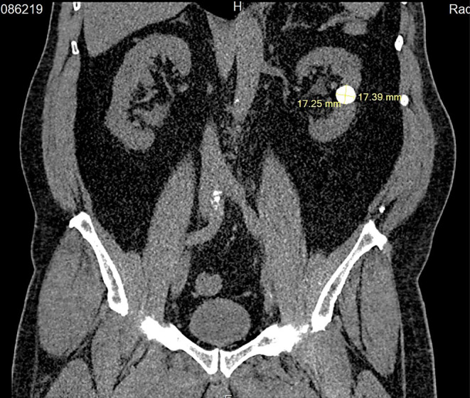
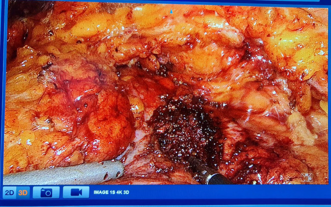
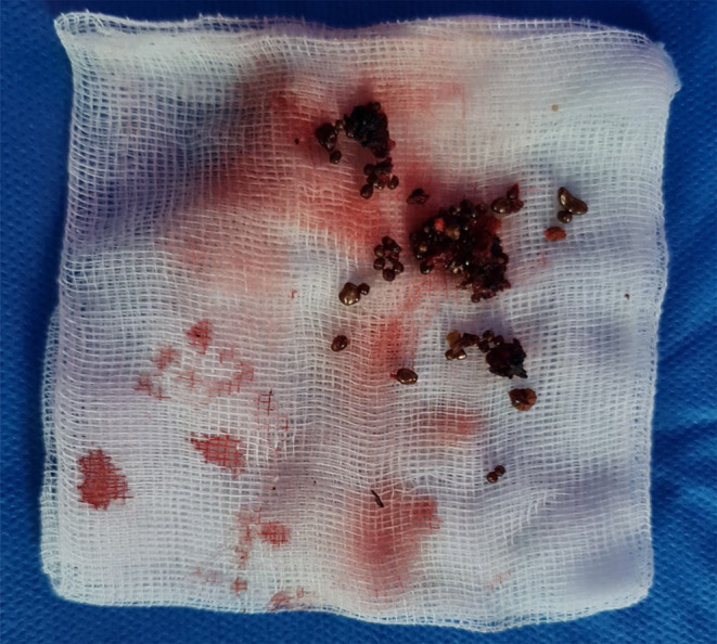
This study aims to present a case of laparoscopic nephrolithotomy and highlight its successful outcome. The patient was a 65-year-old male who experienced intermittent colicky flank pain. Imaging revealed the presence of a 20-mm round-shaped stone in a hydronephrotic calyx located in the mid-pole of the left kidney, possibly with a parenchymal component. Given the stone's location, neither percutaneous nephrolithotripsy (PCNL) nor ureterorenoscopy (URS) was considered feasible. The findings of this study demonstrate the effectiveness and suitability of laparoscopic nephrolithotomy (LNL) in managing stones. It emphasizes that LNL can be utilized not only as an alternative treatment but also as a viable initial option when appropriate case selection is made.
Keywords: case report; laparoscopic nephrolithotomy; nephrolithiasis; struvite stones.
© 2025 The Author(s). Clinical Case Reports published by John Wiley & Sons Ltd.
Conflict of interest statement
The authors declare no conflicts of interest.
Figures
Similar articles
-
[Mini PCNL versus RIRS for renal stones: a prospective randomised controlled study].Urologiia. 2024 Sep;(4):11-15. Urologiia. 2024. PMID: 39563577 Clinical Trial. Russian.
-
The feasibility of ultrasound-guided mini-percutaneous nephrolithotomy for ESWL-resistant lower calyx renal stones of up to two centimeters: a single center experience.J Med Life. 2023 Apr;16(4):520-525. doi: 10.25122/jml-2023-0036. J Med Life. 2023. PMID: 37305831 Free PMC article.
-
An Analysis of Percutaneous Nephrolithotomy (PCNL) Performed at the Institute of Kidney Disease, Pakistan: Stone Clearance and Complications.Cureus. 2024 Dec 9;16(12):e75430. doi: 10.7759/cureus.75430. eCollection 2024 Dec. Cureus. 2024. PMID: 39791059 Free PMC article.
-
Extracorporeal shockwave lithotripsy vs. percutaneous nephrolithotomy vs. flexible ureterorenoscopy for lower-pole stones.Arab J Urol. 2012 Sep;10(3):336-41. doi: 10.1016/j.aju.2012.06.004. Epub 2012 Jul 24. Arab J Urol. 2012. PMID: 26558046 Free PMC article. Review.
-
Endourologic Management (PCNL, URS, SWL) of Stones in Solitary Kidney: A Systematic Review from European Association of Urologists Young Academic Urologists and Uro-Technology Groups.J Endourol. 2020 Jan;34(1):7-17. doi: 10.1089/end.2019.0455. Epub 2019 Sep 26. J Endourol. 2020. PMID: 31456421
References
-
- EAU Guidelines , “Edn. Presented at the EAU Annual Congress Milan 2023,” ISBN 978‐94‐92671‐19‐6.
-
- Stapleton F. B., “Childhood Stones,” Endocrinology and Metabolism Clinics of North America 31 (2002): 1001–1015. [PubMed: 12474642]. - PubMed
LinkOut - more resources
Full Text Sources
