

The benefits of contrast-enhanced ultrasound in the differential diagnosis of suspicious breast lesions
- PMID: 39776839
- PMCID: PMC11703730
- DOI: 10.3389/fmed.2024.1511200
The benefits of contrast-enhanced ultrasound in the differential diagnosis of suspicious breast lesions
Abstract
Background: Contrast-enhanced ultrasound (CEUS) shows potential for the differential diagnosis of breast lesions in general, but its effectiveness remains unclear for the differential diagnosis of lesions highly suspicious for breast cancers.
Objective: This study aimed to evaluate the diagnostic value of CEUS in differentiating pathological subtypes of suspicious breast lesions defined as category 4 of US-BI-RADS.
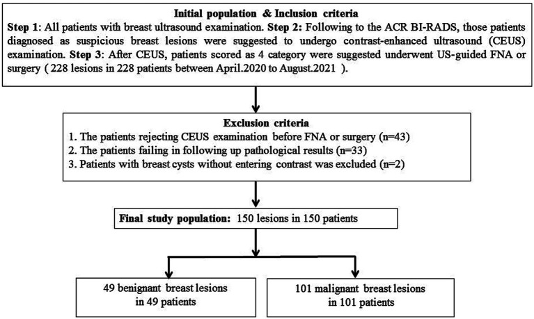
Methods: The dataset of 150 breast lesions was prospectively collected from 150 patients who underwent routine ultrasound and CEUS examination and were highly suspected of having breast cancers. All lesions were pathologically confirmed by US-guided needle biopsy and surgery. The qualitative features and the quantitative parameters of CEUS of these breast lesions were analyzed. The CEUS and biopsy examinations were performed after informed consent.
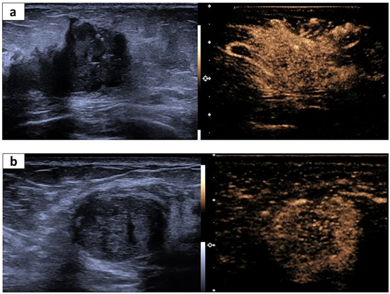
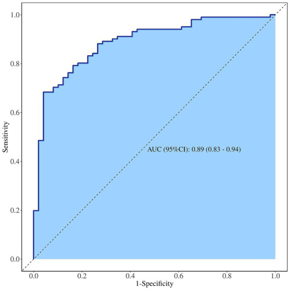
Results: In the qualitative features, crab clam-like enhancement, the presence of more than two enhanced vessels within lesions, and surrounding enriched vessels inserting into lesions were able to differentiate atypical fibroadenomas (FIB) and mass-like non-puerperal mastitis (NPM) from invasive ductal carcinomas (IDC) and ductal carcinomas in situ (DCIS) (p < 0.05). The enlarged scope, irregular shape, and perfusion deficiency were valuable to the differential diagnosis of FIB from the others (p < 0.05). In the four quantitative parameters of CEUS, only the peak intensity (IMAX) contributed to the differential diagnosis between malignant and benign tumors (p < 0.05, ROCAUC: 0.61, sensitivity: 60.4% and specificity: 65.9%, accuracy: 62.1%). However, IMAX did not show any difference in the paired comparison of IDC, DCIS, FIB, and NPM (p > 0.05). The logistic regression analysis results showed that heterogeneous perfusion, crab clam-like enhancement, and partial_ IMAX were independent risk factors for benign and malignant breast lesions (p < 0.05). The area under a receiver operating characteristic of the integrated model was 0.89. In the diagnosis of benign and malignant pathological subtypes of breast lesions, independent risk factors and integrated models had no statistical significance in the diagnosis of IDC and DCISs, FIB, and NPM (p > 0.05).
Conclusion: Some qualitative risk features of CEUS can distinguish malignant breast lesions from NPM and atypical FIB with a high score of US-BI-RADS, aiding physicians to reduce the misdiagnosis of suspicious breast lesions in clinical practice.
Keywords: contrast-enhanced ultrasound; differential diagnosis; qualitative features; quantitative parameter; suspicious breast lesions.
Copyright © 2024 Liang, Lian, Zhang, Jing, Bian, Xu, He, Yu, Zhou and Jiang.
Conflict of interest statement
The authors declare that the research was conducted in the absence of any commercial or financial relationships that could be construed as a potential conflict of interest.
Figures

References
-
- D'Orsi CJ, Sickles E, Mendelson EB. ACR BI-RADS Atlas, Breast imaging reporting and data system. Reston, VA: American College of Radiology; (2013).
LinkOut - more resources
Full Text Sources