Review
. 2024 Nov 22:12:rbae133.
doi: 10.1093/rb/rbae133. eCollection 2025.

Engineered bio-functional material-based nerve guide conduits for optic nerve regeneration: a view from the cellular perspective, challenges and the future outlook

Affiliations
Review

Engineered bio-functional material-based nerve guide conduits for optic nerve regeneration: a view from the cellular perspective, challenges and the future outlook

Enoch Obeng et al. Regen Biomater. .

Abstract

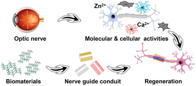
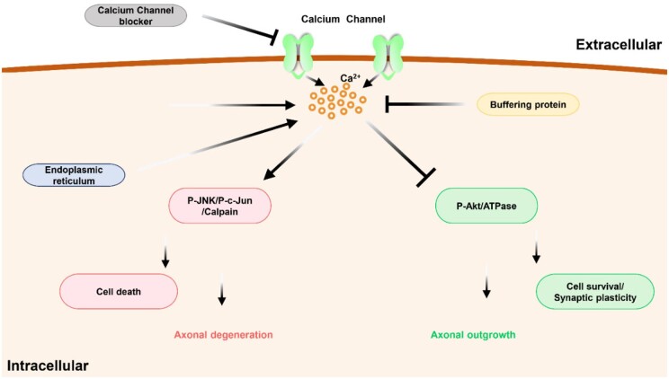
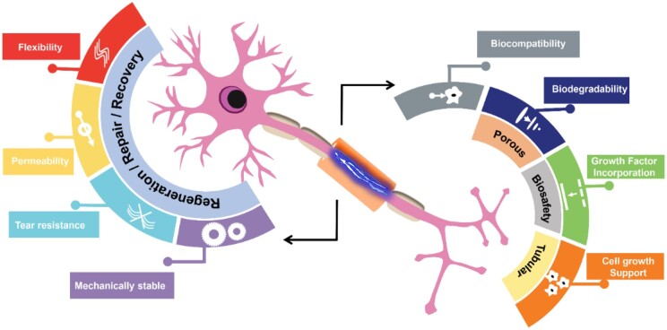
Nerve injuries can be tantamount to severe impairment, standard treatment such as the use of autograft or surgery comes with complications and confers a shortened relief. The mechanism relevant to the regeneration of the optic nerve seems yet to be fully uncovered. The prevailing rate of vision loss as a result of direct or indirect insult on the optic nerve is alarming. Currently, the use of nerve guide conduits (NGC) to some extent has proven reliable especially in rodents and among the peripheral nervous system, a promising ground for regeneration and functional recovery, however in the optic nerve, this NGC function seems quite unfamous. The insufficient NGC application and the unabridged regeneration of the optic nerve could be a result of the limited information on cellular and molecular activities. This review seeks to tackle two major factors (i) the cellular and molecular activity involved in traumatic optic neuropathy and (ii) the NGC application for the optic nerve regeneration. The understanding of cellular and molecular concepts encompassed, ocular inflammation, extrinsic signaling and intrinsic signaling for axon growth, mobile zinc role, Ca2+ factor associated with the optic nerve, alternative therapies from nanotechnology based on the molecular information and finally the nanotechnological outlook encompassing applicable biomaterials and the use of NGC for regeneration. The challenges and future outlook regarding optic nerve regenerations are also discussed. Upon the many approaches used, the comprehensive role of the cellular and molecular mechanism may set grounds for the efficient application of the NGC for optic nerve regeneration.

Keywords: biomaterials; nerve guide conduits; optic nerve crush; optic neuropathy; regeneration.


Figures

None
Graphical abstract
Figure 1.
Figure 1.
Schematic representation of the inflammatory response following ONC.
Figure 2.
Figure 2.
Schematic representation of the yield of A1 and A2 astrocytes via injury and ischemia respectively.
Figure 3.
Figure 3.
Activity of neutrophil in relation to CNTF gene therapy. (A) In situ hybridization detected low levels of CNTFRα mRNA (white) in the RGCs (stained with antibody to RBPMS (red) to delineate RGC cell bodies but not axon bundles). (B) Regenerating axons visualized by CTB immunostaining (green). The asterisk indicates the injury site, and the whole-mounted retinas immunostained with antibody TUJ1+ (green) to visualize βIII tubulin-positive RGCs. (C) Immune cells stained with F4/80 (green) (macrophages), Gr1 (red) and the nuclear marker DAPI (blue). macrophages (Gr1lowF4/80high). (D) Immune cells isolated from blood; stained with fluorescently conjugated antibodies to CD11b, Ly6G and Ly6C. (E) Regenerating axons visualized by CTB immunostaining (green) and its quantification. Retinal whole mounts immunostained for βIII tubulin (antibody TUJ1) (green) 2 weeks after NC and quantitation of RGC survival. With approval, reprinted from Ref. [190]. Copyright 2021, PNAS.
Figure 4.
Figure 4.
Zn2+ Chelation effect on RGC death. (A) Section of flat-mounted retinas immuno-stained (βIII-tubulin, 12 weeks. NC) for visualization of RGCs survival with/without TPEN treatment or pten deletion. (B) Quantitation of long-term RGC survival (C) Intraocular TPEN combined with pten deletion in RGCs for axon regeneration in mice with (D) Longitudinal sections of the mouse optic nerve immuno-stained (GAP-43 2 weeks. pNC) and its quantification. With approval, reprinted from Ref. [261]. Copyright 2017, PNAS.
Figure 5.
Figure 5.
TPEN Effect on RGC survival. (A) Images of GAP-43-immunostained longitudinal sections through the optic nerve 2 weeks after ONC and magnified images of the optic nerve regions proximal and distal to the injury site. (B) Quantitation of axon regeneration 2 weeks after sole and combinatorial. (C) Nuclei marker (DAPI) stained horizontal section through the suprachiasmatic nucleus. Regions of the optic tract with CTB-labeled regenerated axons marked with dotted white lines. Absence of axons at, or growing towards the SCN. (D) Insets of the CTB-labeled axons in the optic tract regions as outlined in (C). With approval, reprinted from Ref [169]. Copyright 2018, Elsevier.
Figure 6.
Figure 6.
Schematic illustration of the inflow of the Ca2+ ions, cell death and axonal degeneration process.
Figure 7.
Figure 7.
Diagrammatic representation of features of a suitable NGC.
Figure 8.
Figure 8.
NGC With some biological cues. (A) Diagrammatic representation of the optic nerve surgery and the release kinetics of CNTF. (B) Images of toluidine blue staining and the transmission electron microscopy of cross-section at the Middle part of the lesion site and the distal optic nerve stump. (C) CTB-labeled RGC axons from the CNTF-chitosan group. (D) The axons in the ventral hypothalamus. (E) The number of the regenerated axons as a function of distance from the proximal stump of the optic nerve. (F) CTB-labeled axons in the SC. High-magnification images of the marked regions. With approval, reprinted from Ref [501]. Copyright 2023, Springer Nature.
Figure 9.
Figure 9.
Function of NGC without biological cues. (A) Pre- and post-heat treatment SEM images of the poly(e-caprolactone) nanofiber wrap. The scaffold has a three-dimensional, interconnected pore structure and consists of randomly oriented fibers. (B) Gross examination of the repair site at 5 weeks post-repair. (C) The denser inflammatory response elicited by the AxoGuard is evidenced by the enormous number of macrophages that invaded its wall as compared to the nanofiber wrap. (D and E) Comparing the number and density of axons in the AxoGuard group as compared to the nanofiber group. With approval, reprinted from Ref [513]. Copyright 2019, Elsevier.
Figure 10.
Figure 10.
Neural cell communication from photoelectric cues. (A) Biocompatibility evaluation of CG_PVA and CG_PVA_Gr electrospun sheets on neuro 2a cells. (B) SEM images of the morphology of neuro 2a cells on (i, ii) CG_PVA, (iii, iv) CG_PVA_Gr nanofibrous sheets on day 3 and day 5 of culture. (C) DAPI staining of neuro 2a cells on (i, ii) CG_PVA, (iii, iv) CG_PVA_Gr nanofibrous sheets. With approval, reprinted from Ref [563]. Copyright 2023, Elsevier.
Figure 11.
Figure 11.
Photocurrent enhanced cell differentiation. (A) Diagrammatic representation of NGCs fabrication and use of PLLA-Ag/Bi2S3 for neural differentiation and nerve regeneration. (B) Representational image of conduit and cell co-culture and (C) cells stained by calcein-AM (green, live cells) and propidium iodide (red, dead cells) after with or without 808 nm NIR irradiation (D) images of PC12 cell differentiation on different substrates. (E) Percentages of differentiated PC12 and average neurite length of differentiated neurons. With approval, reprinted from Ref [573]. Copyright 2022, Elsevier.

Similar articles

References

    1. Hench LL, Polak JM. Third-generation biomedical materials. Science 2002;295:1014–7. - PubMed
    1. Hench LL. Biomaterials. Science 1980;208:826–31. - PubMed
    1. Shirtliff V, Hench L. Bioactive materials for tissue engineering, regeneration and repair. J Mater Sci 2003;38:4697–707.
    1. Hench LL, Xynos ID, Polak JM. Bioactive glasses for in situ tissue regeneration. J Biomater Sci Polym Ed 2004;15:543–62. - PubMed
    1. Iviglia G, Kargozar S, Baino F. Biomaterials, current strategies, and novel nano-technological approaches for periodontal regeneration. J Funct Biomater 2019;10:3. - PMC - PubMed

LinkOut - more resources