Review
. 2024 Nov 5:12:rbae129.
doi: 10.1093/rb/rbae129. eCollection 2025.

Advancements in nanohydroxyapatite: synthesis, biomedical applications and composite developments

Affiliations
Review

Advancements in nanohydroxyapatite: synthesis, biomedical applications and composite developments

Rui Zhao et al. Regen Biomater. .

Abstract

Nanohydroxyapatite (nHA) is distinguished by its exceptional biocompatibility, bioactivity and biodegradability, qualities attributed to its similarity to the mineral component of human bone. This review discusses the synthesis techniques of nHA, highlighting how these methods shape its physicochemical attributes and, in turn, its utility in biomedical applications. The versatility of nHA is further enhanced by doping with biologically significant ions like magnesium or zinc, which can improve its bioactivity and confer therapeutic properties. Notably, nHA-based composites, incorporating metal, polymeric and bioceramic scaffolds, exhibit enhanced osteoconductivity and osteoinductivity. In orthopedic field, nHA and its composites serve effectively as bone graft substitutes, showing exceptional osteointegration and vascularization capabilities. In dentistry, these materials contribute to enamel remineralization, mitigate tooth sensitivity and are employed in surface modification of dental implants. For cancer therapy, nHA composites offer a promising strategy to inhibit tumor growth while sparing healthy tissues. Furthermore, nHA-based composites are emerging as sophisticated platforms with high surface ratio for the delivery of drugs and bioactive substances, gradually releasing therapeutic agents for progressive treatment benefits. Overall, this review delineates the synthesis, modifications and applications of nHA in various biomedical fields, shed light on the future advancements in biomaterials research.

Keywords: bone regeneration; cancer therapy; composite material; dentistry; nanohydroxyapatite.


Figures

None
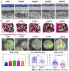
Graphical abstract
Figure 1.
Figure 1.
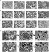
Typical images of nHA crystals. (A) nHA particles by wet chemical precipitation. (B) nHA particles by hydrothermal treatment. (C) nHA particles by heat treatment method. (D) nHA particles by precipitation and treated at 800°C and 1000°C for 2 h.
Figure 2.
Figure 2.
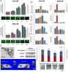
Effect of nHA in osteoporotic bone defects. (A) Cell viability of osteoporotic osteoblasts (OVX-OB) and normal osteoblasts (SHM-OB) in different concentrations of nHA as well as bone sialoprotein (BSP), alkali secretion of osteogenesis-related proteins such as sex phosphatase (ALP) and osteocalcin (OCN). #Significant difference from 0 µg/ml nHA treated SHM-OB group (control) with p < 0.05; *Significant difference from 0 µg/ml nHA treated OVX-OB group (control) with p < 0.05. (B) In vivo evaluation of the distal femurs with drilled defect of OVX and SHM rats, with or without 100 µg/ml nHA treatment after 4 weeks [61]. #Significant difference from SHM-control with p < 0.05; *Significant difference from OVX-control group with p < 0.05. Copyright 2017, Elsevier.
Figure 3.
Figure 3.
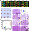
In vivo anti-tumor effects of six inorganic nanoparticles. (A) Thermal images of tumor-bearing mice after a single injection of different inorganic nanoparticles. Three representative mice from each group are shown at day 28. (B) Photographs and (C) the measurement of weight of excised tumors at the end of in vivo studies. *P < 0.05, significant difference was assessed by one-way analysis of variance (ANOVA) followed by Tukey’s post hoc test compared to n-nHA, n-gHA, and n-SiO2 groups. (D) Representative hematoxylin and eosin (H&E) staining images of tumor tissues excised at day 28. Top-right images show tumor tissues at low magnification. Left images show the appearance of nanoparticle aggregates in tumors. Bottom-right images show the highly magnified cells surrounding the nanoparticle aggregates. N, aggregates of nanoparticles. Circles indicate the multinucleated giant cells (MNGCs). (E) Average number of MNGCs counted from 100-fold histology images of tumors representative of three independent mice [13]. *P < 0.05, significant difference was assessed by one-way ANOVA followed by Tukey’s post hoc test compared to n-nHA, n-gHA, and n-SiO2 groups. Ctrl, control. Copyright 2023, Science Advances.
Figure 4.
Figure 4.
Preparation and morphological characterization of various ZQ71 samples. (A) Schematic illustration of the preparation of various ZQ71 samples. (B) Surface SEM images and EDS analysis and (C) cross-section SEM images of various ZQ71 samples [178]. Copyright 2023, Elsevier.
Figure 5.
Figure 5.
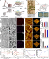
Preparation and characterization of the biomimetic PEKK materials. (A) Schematic illustration of the preparation of biomimetic PEKK scaffold. (B) Regulating effect of the biomimetic PEKK scaffold on osteogenesis and osteoclastogenesis of osteoporotic bone defect. OC, osteoclast; OB, osteoblast. (C) Typical scanning electron microscopy (SEM) and transmission electron microscopy (TEM) images and element mapping of various PEKK scaffolds and natural osteoporotic bone. (D) Atomic force microscopy (AFM) images and corresponding (E) surface roughness of PEKK scaffolds and natural osteoporotic bone. (F) Specific surface area (SSA) of different scaffolds. (G) Water contact angle, (H) Fourier transform infrared spectrophotometer (FTIR) spectra, and (I) X-ray diffractometer (XRD) patterns of different scaffolds. (J) Ca and (K) Sr ion release behavior of different scaffolds in cell culture medium [184]. Copyright 2020, Science Advances.
Figure 6.
Figure 6.
In vivo evaluation of bone healing effect of nHA loaded micro-whiskered hydroxyapatite (nwHA) bioceramics implanted in an osteoporotic rat model with bone defect. (A) Schematic diagram and SEM morphology of micro-nanostructured hydroxyapatite (nwHA) bioceramics. (B) Histological staining analysis and biomechanical test for osteoporotic bone regeneration induced by nwHA bioceramics. (C) Sequential fluorescence labeling of new bone formation inside nwHA bioceramics, with tetracycline and calcein labels shown in distinct fluorescence. Osteogenesis modes include type I osteogenesis(the direction of new bone formation is toward the adjacent hole wall) and type II osteogenesis (the direction of new bone formation is away from adjacent pore walls); comparison of mineral apposition rate (MAR) between different nwHA groups; statistical analysis of the relationship between osteogenesis type and pore diameter of the bioceramics, and the relationship between osteogenesis type and the MAR [234]. Copyright 2023, Elsevier.
Figure 7.
Figure 7.
Perspective key issues regarding the development of nHA and composites.

Similar articles

Cited by

References

    1. Salmasi S, Nayyer L, Seifalian AM, Blunn GW. Nanohydroxyapatite effect on the degradation, osteoconduction and mechanical properties of polymeric bone tissue engineered scaffolds. Open Orthop J 2016;10:900–19. - PMC - PubMed
    1. Yusuf A, Almotairy ARZ, Henidi H, Alshehri OY, Aldughaim MS. Nanoparticles as drug delivery systems: a review of the implication of nanoparticles’ physicochemical properties on responses in biological systems. Polymers (Basel) 2023;15:1596. - PMC - PubMed
    1. Cui L, Wang X, Zhao X, Sun B, Xia T, Hu S. CeO(2) nanoparticles induce pulmonary fibrosis via activating S1P pathway as revealed by metabolomics. Nano Today 2022;45:101559. - PMC - PubMed
    1. Zhang Q, Xiao L, Xiao Y. Porous nanomaterials targeting autophagy in bone regeneration. Pharmaceutics 2021;13:1572. - PMC - PubMed
    1. Bikiaris ND, Koumentakou I, Samiotaki C, Meimaroglou D, Varytimidou D, Karatza A, Kalantzis Z, Roussou M, Bikiaris RD, Papageorgiou GZ. Recent advances in the investigation of poly(lactic acid) (PLA) nanocomposites: incorporation of various nanofillers and their properties and applications. Polymers (Basel) 2023;15:1196. - PMC - PubMed

LinkOut - more resources