. 2024 Dec 3:12:rbae140.
doi: 10.1093/rb/rbae140. eCollection 2025.

FeMOFs/CO loading reduces NETosis and macrophage inflammatory response in PLA based cardiovascular stent materials

Affiliations

FeMOFs/CO loading reduces NETosis and macrophage inflammatory response in PLA based cardiovascular stent materials

Yinhong Xie et al. Regen Biomater. .

Abstract

Modification of polylactic acid (PLA) is a promising strategy for the next generation of bioresorbable vascular stent biomaterials. With this focus, FeMOFs nanoparticles was incorporated in PLA, and then post loading of carbon monoxide (CO) was performed by pressurization. It showed FeMOFs incorporation increased hydrophilicity of the surface and CO loading, and CO release was sustained at least for 3 days. It is well acknowledged NETosis and macrophage mediated inflammation are the principal effectors of atherosclerosis and cardiovascular disease, and it further increases the risk of late stent thrombosis and restenosis. In this study, the effects of CO release of PLA/FeMOFs/CO on NETosis and macrophage behavior were thoroughly explored. In vitro evaluation results showed that PLA/FeMOFs/CO significantly inhibited neutrophil extracellular traps (NETs) release and neutrophil elastase expression by reducing intracellular reactive oxygen species in a simulated inflammatory environment. It reduced Lipopolysaccharide-induced macrophage inflammation with decreased tumor necrosis factor-α expression and increased IL-10 expression. Meanwhile it enhanced endothelial cell activity and growth in inflammatory environment, and inhibited platelet adhesion and activation. In vivo implantation results confirmed that PLA/FeMOFs/CO reduced the macrophages and neutrophils mediated inflammatory response, thus reduced the neointimal hyperplasia. Overall, PLA/FeMOFs/CO effectively prevented the inflammation and restenosis associated with PLA implantation. Our study provides a new strategy to improve the immunocompatibility of PLA implant materials.

Keywords: FeMOFs; NETosis; carbon monoxide; inflammation; macrophage.


Figures

None
Graphical abstract
Figure 1.
Figure 1.
Schematic illustration of PLA/FeMOFs/CO for reducing NETosis and macrophage inflammatory.
Figure 2.
Figure 2.
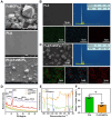
Materials characterization of PLA/FeMOFs/CO. (A) SEM images of FeMOFs nanoparticle, PLA and PLA/FeMOFs. EDS of (B) PLA and (C) PLA/FeMOFs. (D) XRD pattern of FeMOFs nanoparticle, PLA+FeMOFs Co-mingled particles, PLA and PLA/FeMOFs. (E) FT-IR spectra of PLA and PLA/FeMOFs. (F) Water contact angles of PLA and PLA/FeMOFs. Data are presented as means ± SD and analyzed using one-way ANOVA, *P < 0.05, **P < 0.01, ***P < 0.001.
Figure 3.
Figure 3.
CO releasing detected by CO fluorescent probe 1-Ac. (A) Fluorescence spectra of monitor CO release from 1-Ac/CO, FeMOFs/CO, PLA/FeMOFs/CO. (B) Fluorescence spectra of monitor CO release from PLA/FeMOFs/CO after PBS incubation.
Figure 4.
Figure 4.
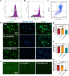
NETosis Inhibition function of PLA/FeMOFs/CO in vitro. (A) FITC-CD11b, (B) PE-Ly6G, (C) FITC-CD11b and PE-Ly6G stains to determine the purity of extracted neutrophils by flow cytometry. (D) Fluorescence images of NETs stained with SYTOX and Hoechst33342 by PMA induced neutrophils. (E) NE and (F) NETs concentration of PMA induced neutrophils quantified by ELISA. (G) Fluorescence images of PMA induced neutrophils stained with DCFH-DA. (H) The quantification results of fluorescence intensity. Data are presented as means ± SD and analyzed using one-way ANOVA, *P < 0.05, **P < 0.01, ***P < 0.001.
Figure 5.
Figure 5.
The anti-inflammatory function of PLA/FeMOFs/CO in vitro. (A) Rhodamine staining of macrophages on samples after 1 day and 3 days culture. (B) CCK-8 assay of macrophages on samples after 1 day and 3 days culture. (C) TNF-α and (D) IL-10 concentration of macrophages quantified by ELISA. Data are presented as means ± SD and analyzed using one-way ANOVA, *P < 0.05, **P < 0.01, ***P < 0.001.
Figure 6.
Figure 6.
HUVECs Growth behavior in simulated inflammatory microenvironment. (A) Rhodamine staining of HUVECs after 24 and 48 h of culture with neutrophil conditioned medium. (B) CCK-8 assay after 24 and 48 h of culture with neutrophil conditioned medium. (C) Rhodamine staining of HUVECs after 24 and 48 h of culture with macrophage conditioned medium. (D) CCK-8 assay after 24 and 48 h of culture with macrophage conditioned medium. Data are presented as means ± SD and analyzed using one-way ANOVA, *P < 0.05, **P < 0.01, ***P < 0.001.
Figure 7.
Figure 7.
In vitro hemocompatibility evaluation. (A) Hemolysis rate of PLA and PLA/FeMOFs. (B) SEM observation of the platelet adhesion and activation. (C) Platelet adhesion level after 30 min incubation with PRP. Data are presented as means ± SD and analyzed using one-way ANOVA, *P < 0.05, **P < 0.01, ***P < 0.001.
Figure 8.
Figure 8.
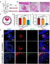
In vivo Sprague-Dawley rats implantation experiments. (A) In vivo implantation rat model. (B) Hematoxylin and eosin-stained after implantation for 30 days. (C) Schematic diagram of neointimal thickness measurement. (D) The quantification results of the neointimal tissue thickness. (E) Images of CD68 immunofluorescence, CD163 immunofluorescence and nuclei were stained with DAPI. (F) Statistics of CD68 fluorescence intensity in neointimal tissue. Data are presented as means ± SD and analyzed using one-way ANOVA, *P < 0.05, **P < 0.01, ***P < 0.001.
Figure 9.
Figure 9.
Evaluation of neutrophil infiltration and endothelialization in vivo. (A) Images of MPO immunofluorescence, and nuclei were stained with DAPI. (B) Statistics of MPO fluorescence intensity in neointimal tissue. (C) Images of CD31 immunofluorescence. The nuclei were stained with DAPI. (D) Quantification of endothelialization. Data are presented as means ± SD and analyzed using one-way ANOVA, *P < 0.05, **P < 0.01, ***P < 0.001.

Similar articles

Cited by

References

    1. Tsao CW, Aday AW, Almarzooq ZI, Alonso A, Beaton AZ, Bittencourt MS, Boehme AK, Buxton AE, Carson AP, Commodore-Mensah Y, Elkind MSV, Evenson KR, Eze-Nliam C, Ferguson JF, Generoso G, Ho JE, Kalani R, Khan SS, Kissela BM, Knutson KL, Levine DA, Lewis TT, Liu J, Loop MS, Ma J, Mussolino ME, Navaneethan SD, Perak AM, Poudel R, Rezk-Hanna M, Roth GA, Schroeder EB, Shah SH, Thacker EL, VanWagner LB, Virani SS, Voecks JH, Wang N-Y, Yaffe K, Martin SS. Heart disease and stroke statistics—2022 update: a report from the American Heart Association. Circulation 2022;145:e153–e639. - PubMed
    1. Scafa Udriște A, Niculescu A-G, Grumezescu AM, Bădilă E. Cardiovascular stents: a review of past, current, and emerging devices. Materials 2021;14:2498. - PMC - PubMed
    1. Schapiro-Dufour E, Tricotel A, Slama MS, Ducimetière P, Trinh-Duc A, Sichel C, Le Tri T, Galmiche H, Dray-Spira R, Zureik M. Major ischaemic and bleeding risks following current drug-eluting stent implantation: are there differences across current drug-eluting stent types in real life? Arch Cardiovasc Dis 2019;112:469–84. - PubMed
    1. Dworeck C, Angeras O, Haraldsson I, Ioanes D, Odenstedt J, Petursson P, Robertsson L, Stewart J, Volz S, Albertsson P, Råmunddal T, Omerovic E. TCT-477 long-term risk of stent thrombosis and restenosis after treatment with drug-eluting stents: a report from SCAAR. J Am Coll Cardiol 2016;68:B191–2.
    1. Lasprilla AJR, Martinez GAR, Lunelli BH, Jardini AL, Filho RM. Poly-lactic acid synthesis for application in biomedical devices–a review. Biotechnol Adv 2012;30:321–8. - PubMed

LinkOut - more resources