

Single-cell Atlas reveals core function of CPVL/MSR1 expressing macrophages in the prognosis of triple-negative breast cancer
- PMID: 39776914
- PMCID: PMC11703973
- DOI: 10.3389/fimmu.2024.1501009
Single-cell Atlas reveals core function of CPVL/MSR1 expressing macrophages in the prognosis of triple-negative breast cancer
Abstract
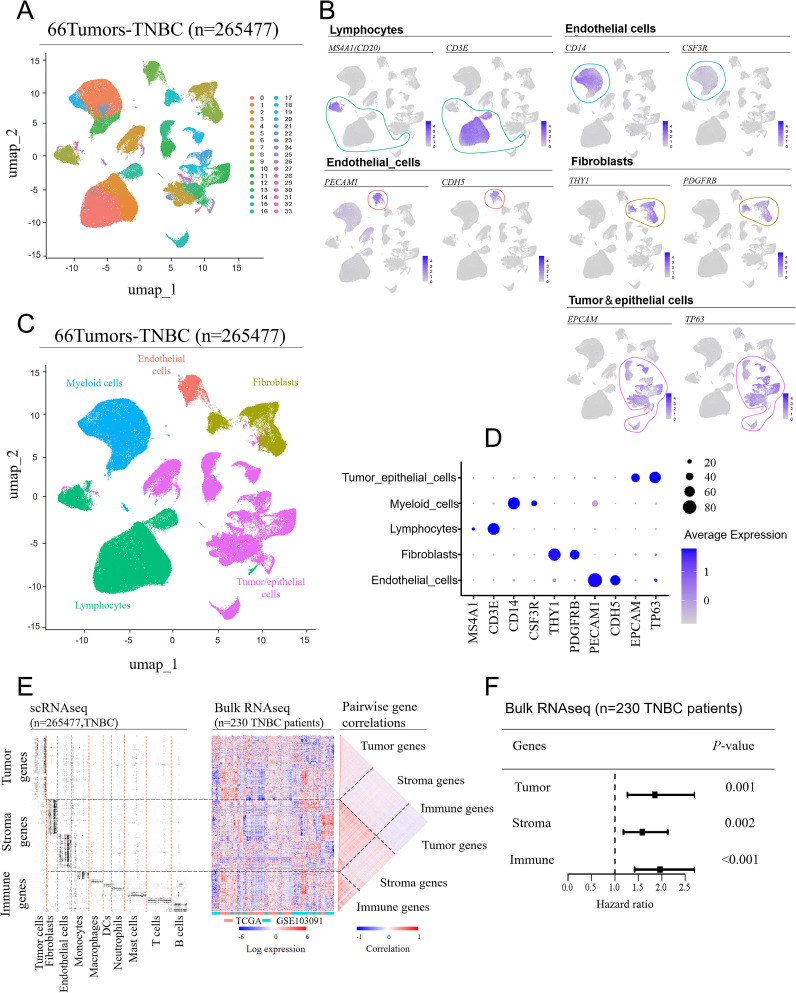
Background: Triple-negative breast cancer (TNBC) is the most aggressive subtype of breast cancer, with the worst prognosis among all subtypes. The impact of distinct cell subpopulations within the tumor microenvironment (TME) on TNBC patient prognosis has yet to be clarified.
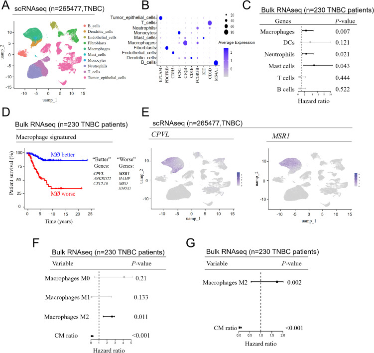
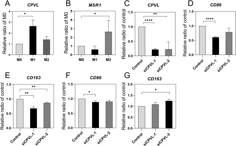
Methods: Utilizing single-cell RNA sequencing (scRNA-seq) integrated with bulk RNA sequencing (bulk RNA-seq), we applied Cox regression models to compute hazard ratios, and cross-validated prognostic scoring using a GLMNET-based Cox model. Cell communication analysis was used to elucidate the potential mechanisms of CPVL and MSR1. Ultimately, RNA interference-mediated gene knockdown was utilized to validate the impact of specific genes on the polarization of tumor-associated macrophages (TAMs).
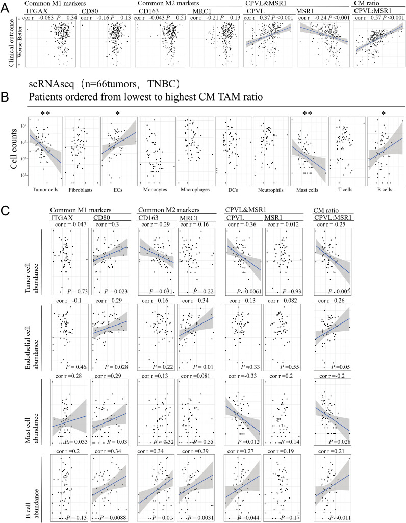
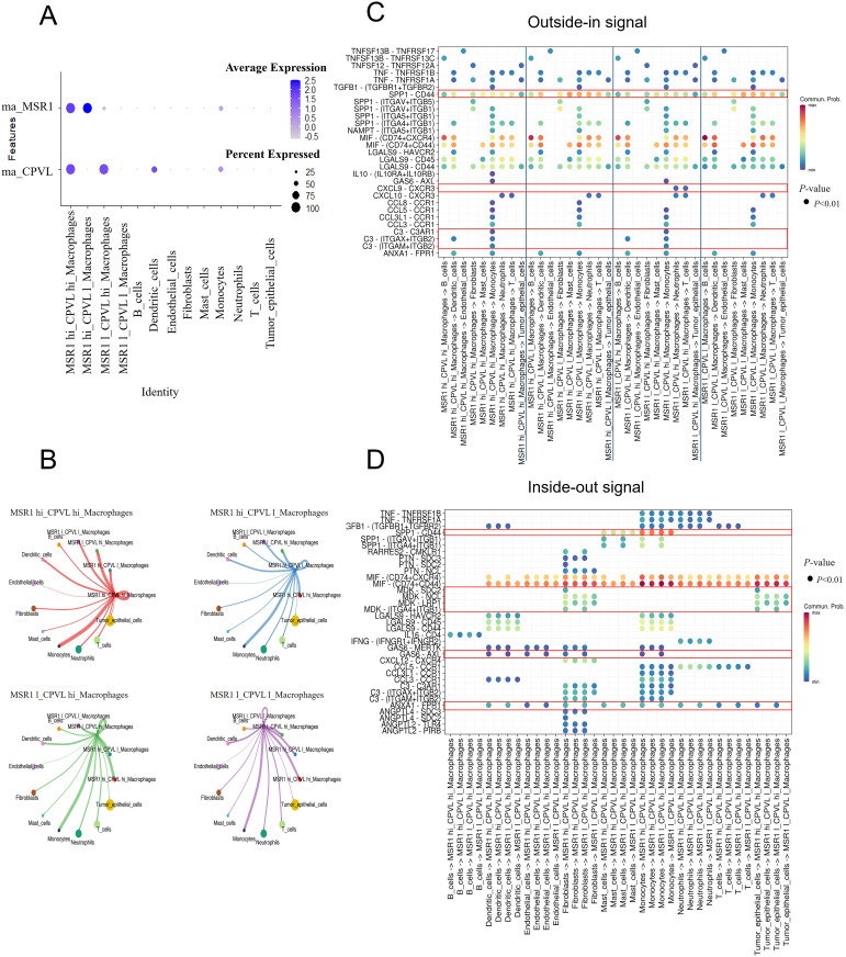
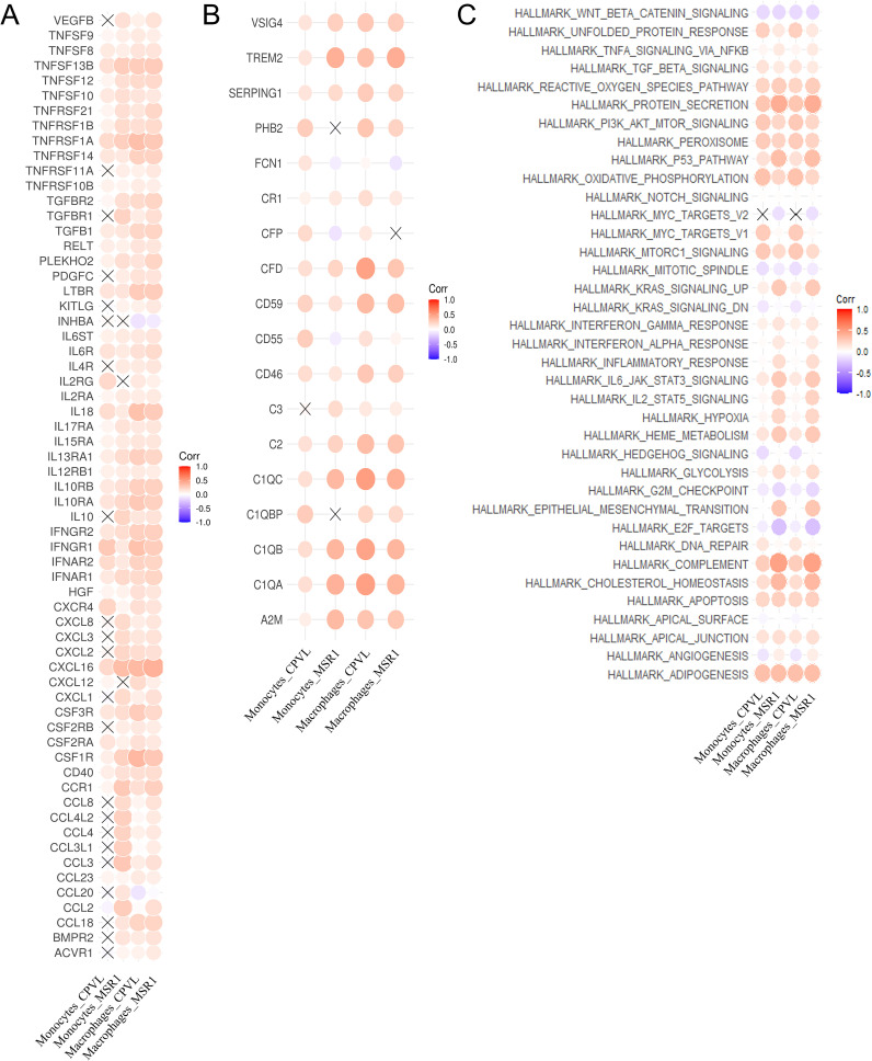
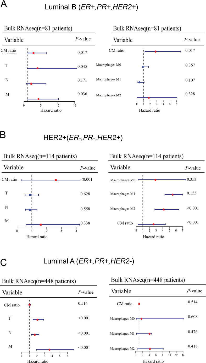
Results: Our findings revealed that the function of immune cells is more pivotal in prognosis, with TAMs showing the strongest correlation with TNBC patient outcomes, compared with other immune cells. Additionally, we identified CPVL and MSR1 as critical prognostic genes within TAMs, with CPVL expression positively correlated with favorable outcomes and MSR1 expression associated with poorer prognosis. Mechanistically, CPVL may contribute to favorable prognosis by inhibiting the SPP1-CD44 ligand-receptor and promoting CXCL9-CXCR3, C3-C3AR1 ligand-receptor, through which TAMs interact with other cells such as monocytes, neutrophils, and T cells. Moreover, cytokines including IL-18, IFNγR1, CCL20, and CCL2, along with complement-related gene like TREM2 and complement component CFD, may participate in the process of CPVL or MSR1 regulating macrophage polarization. Furthermore, RT-PCR experiments confirmed that CPVL is positively associated with M1-like TAM polarization, while MSR1 is linked to M2-like TAM polarization. Finally, the prognostic significance of these two genes is also validated in HER2-positive breast cancer subtypes.
Conclusions: CPVL and MSR1 are potential biomarkers for macrophage-mediated TNBC prognosis, suggesting the therapeutic potential of macrophage targeting in TNBC.
Keywords: MSR1; cPVL; macrophages; prognosis; single-cell sequence; triple-negative breast cancer.
Copyright © 2024 Wang, Lin, Zhang, Zhang, Sun, Yang, Zhang, Yuan, Zhang, Chen and Wen.
Conflict of interest statement
The authors declare that the research was conducted in the absence of any commercial or financial relationships that could be construed as a potential conflict of interest.
Figures
Similar articles
-
Tumor-expressed GPNMB orchestrates Siglec-9+ TAM polarization and EMT to promote metastasis in triple-negative breast cancer.Proc Natl Acad Sci U S A. 2025 Sep 9;122(36):e2503081122. doi: 10.1073/pnas.2503081122. Epub 2025 Sep 2. Proc Natl Acad Sci U S A. 2025. PMID: 40892920
-
Programmed cell death-related prognostic genes mediate dysregulation of the immune microenvironment in triple-negative breast cancer.Front Immunol. 2025 Mar 12;16:1563630. doi: 10.3389/fimmu.2025.1563630. eCollection 2025. Front Immunol. 2025. PMID: 40145099 Free PMC article.
-
Construction of a stromal cell-related prognostic signature based on a 101-combination machine learning framework for predicting prognosis and immunotherapy response in triple-negative breast cancer.Front Immunol. 2025 May 14;16:1544348. doi: 10.3389/fimmu.2025.1544348. eCollection 2025. Front Immunol. 2025. PMID: 40438115 Free PMC article.
-
Macrophages: Key Players in the Battle against Triple-Negative Breast Cancer.Int J Mol Sci. 2024 Oct 7;25(19):10781. doi: 10.3390/ijms251910781. Int J Mol Sci. 2024. PMID: 39409110 Free PMC article. Review.
-
Triple negative breast cancer: Key role of Tumor-Associated Macrophages in regulating the activity of anti-PD-1/PD-L1 agents.Biochim Biophys Acta Rev Cancer. 2018 Jan;1869(1):78-84. doi: 10.1016/j.bbcan.2017.10.007. Epub 2017 Nov 7. Biochim Biophys Acta Rev Cancer. 2018. PMID: 29126881 Review.
References
-
- Guo S, Liu X, Zhang J, Huang Z, Ye P, Shi J, et al. . Integrated analysis of single-cell RNA-seq and bulk RNA-seq unravels T cell-related prognostic risk model and tumor immune microenvironment modulation in triple-negative breast cancer. Comput Biol Med. (2023) 161:107066. doi: 10.1016/j.compbiomed.2023.107066 - DOI - PubMed
MeSH terms
Substances
LinkOut - more resources
Full Text Sources
Research Materials
Miscellaneous