

Clinical, laboratory, and orbital imaging features of giant cell arteritis in comparison to non-arteritic anterior ischemic optic neuropath: a single center case series
- PMID: 39776976
- PMCID: PMC11703802
- DOI: 10.3389/fopht.2024.1498968
Clinical, laboratory, and orbital imaging features of giant cell arteritis in comparison to non-arteritic anterior ischemic optic neuropath: a single center case series
Abstract
Background: Giant cell arteritis (GCA) is the most common vasculitis in patients older than 50 years and is considered a "do not miss" diagnosis. However, it remains a diagnostic challenge given overlapping clinical syndromes such as non-arteritic anterior ischemic optic neuropathy (NAION) and poorly explored imaging findings.
Materials and methods: In this retrospective study between the time period of January 2013 and December 2021, a total of 13 consecutive patients with a pathological diagnosis of GCA and 8 patients with clinical diagnosis of NAION were isolated. Demographic and clinical data for each patient were collected, including pertinent laboratory data. Pertinent physical exam data was also collected, including fundoscopic exam and visual acuity. Two neuroradiologist assessed the orbital MRI imaging findings of GCA and NAION for the presence and characterization of imaging abnormalities. Assessment for potential relationship between GCA orbital findings, laboratory and visual outcomes was performed. Finally, comparison between GCA and NAION imaging findings was performed.
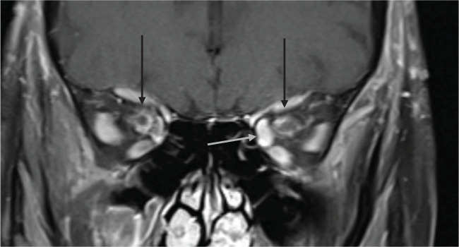
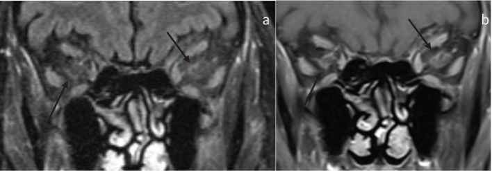
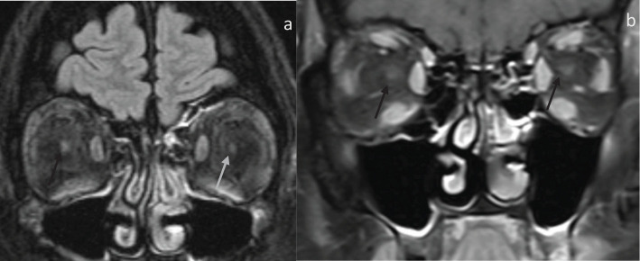
Results: 13 GCA patients were assessed. 9 patients had abnormal orbital findings. Of these 8 patients had bilateral orbital involvement The most common imaging findings was perineuritis of the optic nerve sheath, present in 7 patients. In total, 8 NAION patients were assessed. All patients demonstrate optic nerve involvement. The Snellen test was converted to logmar, and visual acuity was assessed for both NAION and GCA for each eye at diagnosis and at the last follow-up. There was no statistical significance for either eye for both GCA and NAION at initial diagnosis and final follow-up. In the 4 GCA patients with normal MRI findings and 9 GCA patients with abnormal MRI findings, there was no statistical significance between initial presentation and final follow-up visual acuity.
Conclusion: GCA and NAION are potentially overlapping clinical syndromes with different treatment approach and poorly explored imaging findings. Our case series assesses the orbital imaging findings of both syndromes while noting different imaging pattern of both on MRI, which can serve as a potential tool to aid in diagnosis of both. .
Keywords: MRI; NAION; giant cell (temporal) arteritis; optic nerve (ON); optic neuritis (ON).
Copyright © 2024 Eldaya, Yeh, Stunkel, Parsons and Van Stavern.
Conflict of interest statement
The authors declare that the research was conducted in the absence of any commercial or financial relationships that could be construed as a potential conflict of interest. The author(s) declared that they were an editorial board member of Frontiers, at the time of submission. This had no impact on the peer review process and the final decision.
Figures
Similar articles
-
Incidence of giant cell arteritis mimicking non-arteritic anterior optic neuropathy.J Neurol Sci. 2023 Jun 15;449:120661. doi: 10.1016/j.jns.2023.120661. Epub 2023 Apr 23. J Neurol Sci. 2023. PMID: 37126919
-
Optic nerve sheath enhancement on orbital MRI in giant cell arteritis.Br J Ophthalmol. 2025 May 30;109(6):709-714. doi: 10.1136/bjo-2024-326608. Br J Ophthalmol. 2025. PMID: 39694603
-
Arteritic Anterior Ischemic Optic Neuropathy Associated with Giant-Cell Arteritis in Korean Patients: A Retrospective Single-Center Analysis and Review of the Literature.J Clin Neurol. 2019 Jul;15(3):386-392. doi: 10.3988/jcn.2019.15.3.386. J Clin Neurol. 2019. PMID: 31286712 Free PMC article.
-
Orbital magnetic resonance imaging of giant cell arteritis with ocular manifestations: a systematic review and individual participant data meta-analysis.Eur Radiol. 2023 Nov;33(11):7913-7922. doi: 10.1007/s00330-023-09770-2. Epub 2023 May 31. Eur Radiol. 2023. PMID: 37256352 Free PMC article.
-
Arteritic anterior ischaemic optic neuropathy: An update.Eur J Ophthalmol. 2021 Nov;31(6):2818-2827. doi: 10.1177/11206721211009447. Epub 2021 Apr 23. Eur J Ophthalmol. 2021. PMID: 33892603 Review.
References
-
- Diamantopoulos AP, Haugeberg G, Lindland A, Myklebust G. The fast-track ultrasound clinic for early diagnosis of giant cell arteritis significantly reduces permanent visual impairment: towards a more effective strategy to improve clinical outcome in giant cell arteritis? Rheumatol (Oxford). (2016) 55:66–70. doi: 10.1093/rheumatology/kev289 - DOI - PubMed
LinkOut - more resources
Full Text Sources
Miscellaneous