. 2024 Dec 19;22(2):34.
doi: 10.3892/br.2024.1912. eCollection 2025 Feb.

Bioactive potency of extracts from Stylissa carteri and Amphimedon chloros with silver nanoparticles against cancer cell lines and pathogenic bacteria

Affiliations

Bioactive potency of extracts from Stylissa carteri and Amphimedon chloros with silver nanoparticles against cancer cell lines and pathogenic bacteria

Moath Alqaraleh et al. Biomed Rep. .

Abstract

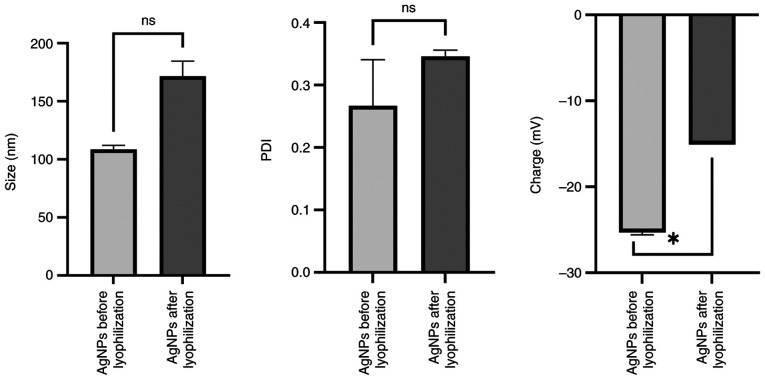
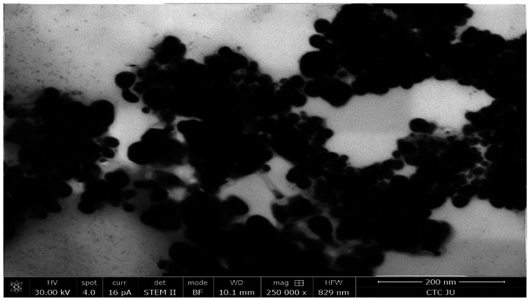
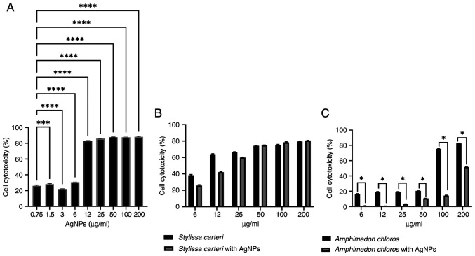
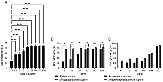
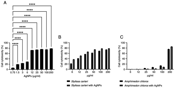
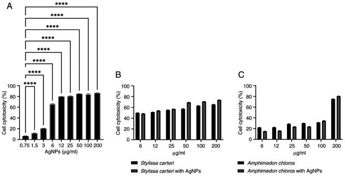
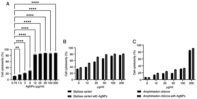
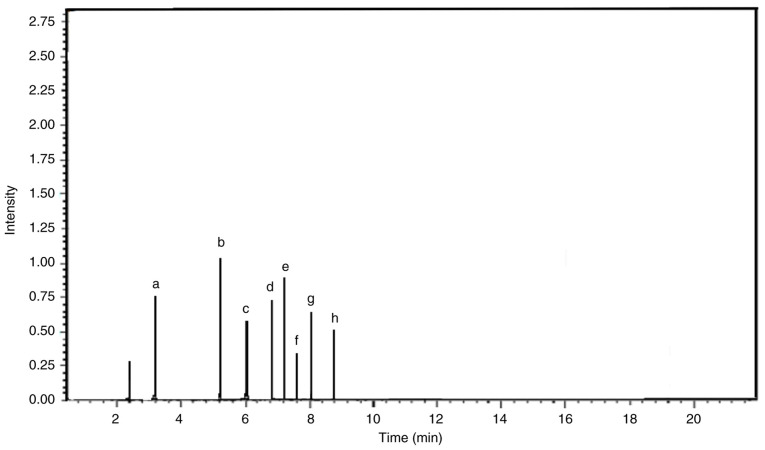
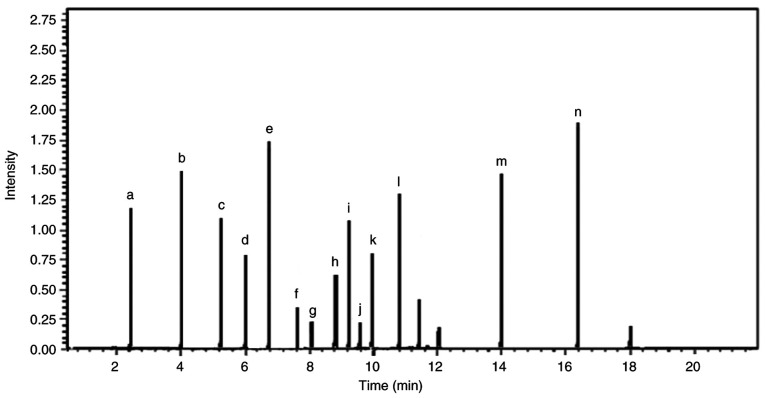
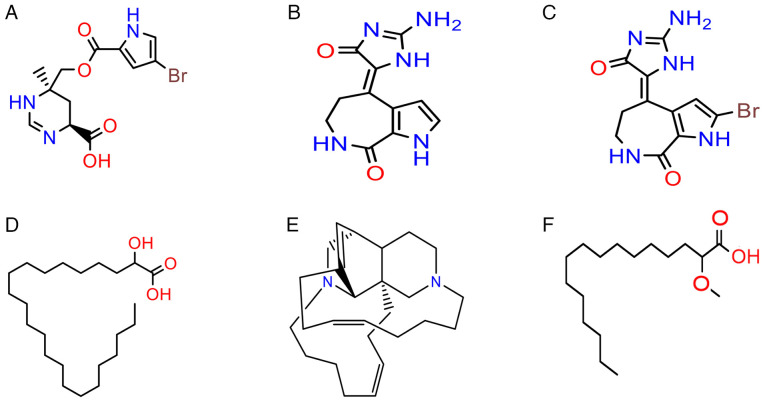
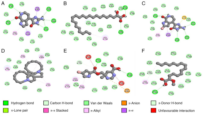
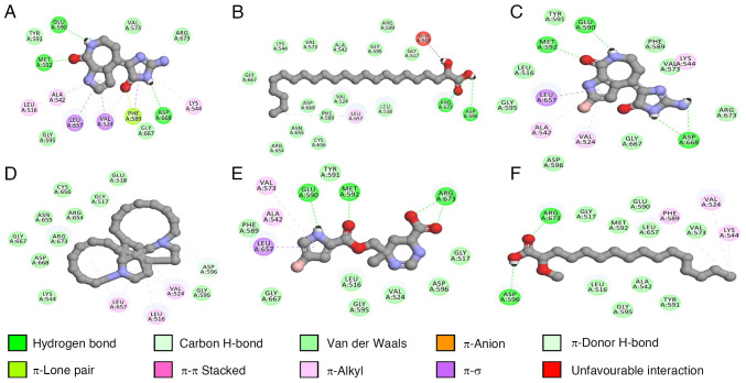
Silver nanoparticles (AgNPs) are spherical particles with a number of specific and unique physical (such as surface plasmon resonance, high electrical conductivity and thermal stability) as well as chemical (including antimicrobial activity, catalytic efficiency and the ability to form conjugates with biomolecules) properties. These properties allow AgNPs to exhibit desired interactions with the biological system and make them prospective candidates for use in antibacterial and anticancer activities. AgNPs have a quenching capacity, which produces reactive oxygen species and disrupts cellular processes (such as reducing the function of the mitochondria, damaging the cell membrane, inhibiting DNA replication and altering protein synthesis). In addition, sponge extracts contain biologically active substances with therapeutic effects. Therefore, the concurrent use of these agents may present a potential for the development of novel antitumor and antimicrobial drugs. The present study investigated the cytotoxic effects of AgNPs combined with the extracts from sponge species, Stylissa carteri or Amphimedon chloros, against various cancer cell lines and pathogenic bacterial strains. The present study was novel as it provided a further understanding of the cytotoxicity and underlying mechanisms of AgNPs. Alterations in the properties, such as size, charge and polydispersity index, of the AgNPs were demonstrated after lyophilization. Scanning electron microscopy revealed submicron-sized particles. The cytotoxic potential of AgNPs across various cancer cell lines such as lung, colorectal, breast and pancreatic cancer cell lines, was demonstrated, especially when the AgNPs were combined with sponge extracts, which suggested a synergistic effect. Analysis using liquid chromatography-mass spectrometry revealed key chemical components in the extracts, and molecular docking simulations indicated potential inhibition interactions between a number of the extract components and the epidermal growth factor receptor and tyrosine kinase receptor A. Synergistic antibacterial effects against several bacterial species such as Staphylococcus xylosus, Klebsiella oxytoca, Enterobacter aerogenes, Micrococcus spp. and Escherichia coli, were observed when AgNPs were combined with sponge ethyl acetate extracts. The results of the present study suggested a potential therapeutic application of marine-derived compounds and nanotechnology in combating cancer and bacterial infections. Future research should further elucidate the mechanistic pathways and investigate the in vivo therapeutic efficacy.

Keywords: AgNPs; Amphimedon chloros; EGFR inhibition; Stylissa carteri; TrkA inhibition; antibacterial activity.


Conflict of interest statement

The authors declare that they have no competing interests.

Figures

Figure 1
Figure 1
Size, PDI and surface charge of AgNPs before and after lyophilization. While there was not a significant difference in the size and PDI of the AgNPs before and after lypophilization, a significant difference in the surface charge was revealed after lyophilization. The results are presented as means ± SD (n=3). *P<0.05. AgNPs, silver nanoparticles; PDI, polydispersity index; ns, not significant.
Figure 2
Figure 2
SEM micrograph of AgNPs. A high-resolution SEM image of the morphology and distribution of AgNPs, which were synthesized using 1.0 mM silver nitrate and the fungus Aspergillus flavus. SEM, scanning electron microscopy; AgNPs, silver nanoparticles; HV, high voltage (accelerating voltage in kV applied to the microscope); spot, spot size (beam diameter for optimal resolution); pA, picoamperes; curr, current (electron beam current in pA); det, detector (type of detector used to capture the image); STEM II, scanning transmission electron microscopy secondary image; BF, bright field (imaging mode providing high-contrast details in transmission images); WD, working distance (distance between sample surface and objective lens); mag, magnification; HFW, horizontal field width; CTC JU, Cells Therapy Center, Jordan University.
Figure 3
Figure 3
Cytotoxicity of HUVEC cells to AgNPs, Stylissa carteri and Amphimedon chloros. (A) Cytotoxicity of different concentrations of AgNPs on the HUVEC cell line (analyzed using one-way ANOVA followed by the Dunnett's test). Cytotoxicity of (B) the Stylissa carteri extract alone and in combination with 0.75 µg/ml AgNPs and (C) the Amphimedon chloros extract alone and in combination with 0.75 µg/ml AgNPs on the endothelial HUVEC cell line (analyzed using unpaired t-tests). The results are presented as means ± SD (n=3). *P<0.05, ***P<0.001 and ****P<0.0001. AgNPs, silver nanoparticles; HUVEC, human umbilical vein endothelial cell line.
Figure 4
Figure 4
Cytotoxicity of A549 cells to AgNPs, Stylissa carteri and Amphimedon chloros. (A) Cytotoxicity of different concentrations of AgNPs on the lung cancer A549 cell line (analyzed using one-way ANOVA followed by the Dunnett's test). Cytotoxicity of (B) the Stylissa carteri extract alone and in combination with 0.75 µg/ml AgNPs and (C) the Amphimedon chloros extract alone and in combination with 0.75 µg/ml AgNPs on the lung cancer A549 cell line (analyzed using unpaired t-tests). The results are presented as means ± SD (n=3). *P<0.05 and ****P<0.0001. AgNPs, silver nanoparticles.
Figure 5
Figure 5
Cytotoxicity of MCF7 cells to AgNPs, Stylissa carteri and Amphimedon chloros. (A) Cytotoxicity of different concentrations of AgNPs on the breast cancer MCF7 cell line (analyzed using one-way ANOVA followed by the Dunnett's test). Cytotoxicity of (B) the Stylissa carteri extract alone and in combination with 0.75 µg/ml AgNPs and (C) the Amphimedon chloros extract alone and in combination with 0.75 µg/ml AgNPs on the breast cancer MCF7 cell line (analyzed using unpaired t-tests). The results are presented as means ± SD (n=3). ****P<0.0001. AgNPs, silver nanoparticles.
Figure 6
Figure 6
Cytotoxicity of PANC-1 cells to AgNPs, Stylissa carteri and Amphimedon chloros. (A) Cytotoxicity of different concentrations of AgNPs on the pancreatic cancer PANC-1 cell line (analyzed using one-way ANOVA followed by the Dunnett's test). Cytotoxicity of (B) the Stylissa carteri extract alone and in combination with 0.75 µg/ml AgNPs and (C) the Amphimedon chloros extract alone and in combination with 0.75 µg/ml AgNPs on the pancreatic cancer PANC-1 cell line (analyzed using unpaired t-tests). The results are presented as means ± SD (n=3). ****P<0.0001. AgNPs, silver nanoparticles.
Figure 7
Figure 7
Cytooxicity of HT-29 cells to AgNPs, Stylissa carteri and Amphimedon chloros. (A) Cytotoxicity of different concentrations of AgNPs on the colorectal cancer HT-29 cell line (analyzed using one-way ANOVA followed by the Dunnett's test). Cytotoxicity of (B) the Stylissa carteri extract alone and in combination with 0.75 µg/ml AgNPs and (C) the Amphimedon chloros extract alone and in combination with 0.75 µg/ml AgNPs on the colorectal cancer HT-29 cell line (analyzed using unpaired t-tests). The results are presented as means ± SD (n=3). **P<0.01 and ****P<0.0001. AgNPs, silver nanoparticles.
Figure 8
Figure 8
Liquid chromatography-mass spectrometry chromatogram of the components in the Stylissa carteri extract. Chromatogram of the Stylissa carteri extract with peaks that correspond to individual compounds, which were identified based on their mass-to-charge ratio. Each peak provided detail on the chemical composition of the extract and the relative abundance of its components. The chromatogram revealed the molecular profile of Stylissa carteri and highlighted key compounds such as (a) debromohymenialdisine, (b) hymenialdisine, (c) agelongine, (d) calthramide, (e) manzacidine A, (f) 3-bromohymenialdisine, (g) spongiacidine and (h) ageliferin. The chromatogram corresponds to ions with an m/z of 193.00, which have a relative intensity of 81.82% compared with the total ion intensity across all detected m/z values. The maximum intensity range or full-scale limit of the detector was 1,000,000,000. m/z, mass-to-charge ratio.
Figure 9
Figure 9
Liquid chromatography-mass spectrometry chromatogram of the components in the Amphimedon chloros extract. Chromatogram of the Amphimedon chloros extract with peaks that correspond to individual compounds, which were identified based on their mass-to-charge ratio. Each peak provided detail on the chemical composition of the extract and the relative abundance of its components. The chromatogram revealed the molecular profile of Amphimedon chloros and highlighted key compounds such as (a) purine, (b) methoxyhexadecanoic acid, (c) hachijodine, (d) tricosenal, (e) kermaphidine B, (f) tricosenoic acid, (g) pentacosenal, (h) zamamidine, (i) karamamine, (j) pentacosenic acid, (k) ircinol, (l) pyrinodemin, (m) nakinadine and (n) hydroxytricosanoic acid. The chromatogram corresponds to ions with an m/z of 193.00, which have a relative intensity of 81.82% compared with the total ion intensity across all detected m/z values. The maximum intensity range or full-scale limit of the detector was 1,000,000,000. m/z, mass-to-charge ratio.
Figure 10
Figure 10
Chemical structures of the main components in the Stylissa carteri and Amphimedon chloros extracts. (A) Manzacidine A, (B) debromohymenialdisine and (C) hymenialdisine were the main components in the Stylissa carteri extract. (D) Hydroxytricosanoic acid, (E) kermaphidine B and (F) methoxyhexadecanoic acid were the main components in the Amphimedon chloros extract.
Figure 11
Figure 11
Binding interactions of six marine-derived compounds with the epidermal growth factor receptor. Ball and stick representation of (A) debromohymenialdisine, (B) hydroxytricosanoic acid, (C) hymenialdisine, (D) keramaphidin B, (E) manzacidine A and (F) methoxyhexadecanoic acid docked against the epidermal growth factor receptor kinase domain (Protein Data Bank ID, 4I23; https://www.rcsb.org/structure/4i23). The figure was created using Biovia Discovery Studio Visualiser 2016® (Dassault Systèmes). H-bond, hydrogen bond.
Figure 12
Figure 12
Binding interactions of six marine-derived compounds with the tyrosine kinase receptor A. Ball and stick representation of (A) debromohymenialdisine, (B) hydroxytricosanoic acid, (C) hymenialdisine, (D) keramaphidin B, (E) manzacidine A and (F) methoxyhexadecanoic acid docked against tyrosine kinase receptor A (Protein Data Bank ID, 7VKO; https://www.rcsb.org/structure/7VKO). The figure was created using Biovia Discovery Studio Visualiser 2016® (Dassault Systèmes). H-bond, hydrogen bond.

Similar articles

References

    1. Gerstberger S, Jiang Q, Ganesh KJC. Metastasis. Cell. 2023;186:1564–1579. doi: 10.1016/j.cell.2023.03.003. - DOI - PMC - PubMed
    1. Siegel RL, Miller KD, Wagle NS, Jemal A. Cancer statistics, CA Cancer J. Clin. 2023;73:17–48. doi: 10.3322/caac.21763. - DOI - PubMed
    1. Bazeed AY, Day CM, Garg SJC. Pancreatic cancer: Challenges and opportunities in locoregional therapies. Cancers (Basel) 2022;14(4257) doi: 10.3390/cancers14174257. - DOI - PMC - PubMed
    1. Vasan N, Baselga J, Hyman DM. A view on drug resistance in cancer. Nature. 2019;575:299–309. doi: 10.1038/s41586-019-1730-1. - DOI - PMC - PubMed
    1. Hossain CM, Gera M, Ali KA. Current status and challenges of herbal drug development and regulatory aspect: A global perspective. Asian J Pharmaceutical Clin Res. 2022;15:31–41.

LinkOut - more resources