. 2024 Dec 24:15:1516513.
doi: 10.3389/fgene.2024.1516513. eCollection 2024.

Urinary TYROBP and HCK as genetic biomarkers for non-invasive diagnosis and therapeutic targeting in IgA nephropathy

Affiliations

Urinary TYROBP and HCK as genetic biomarkers for non-invasive diagnosis and therapeutic targeting in IgA nephropathy

Boji Xie et al. Front Genet. .

Abstract

Background: IgA nephropathy (IgAN) is a leading cause of renal failure, but its pathogenesis remains unclear, complicating diagnosis and treatment. The invasive nature of renal biopsy highlights the need for non-invasive diagnostic biomarkers. Bulk RNA sequencing (RNA-seq) of urine offers a promising approach for identifying molecular changes relevant to IgAN.

Methods: We performed bulk RNA-seq on 53 urine samples from 11 untreated IgAN patients and 11 healthy controls, integrating these data with public renal RNA-seq, microarray, and scRNA-seq datasets. Machine learning was used to identify key differentially expressed genes, with protein expression validated by immunohistochemistry (IHC) and drug-target interactions explored via molecular docking.

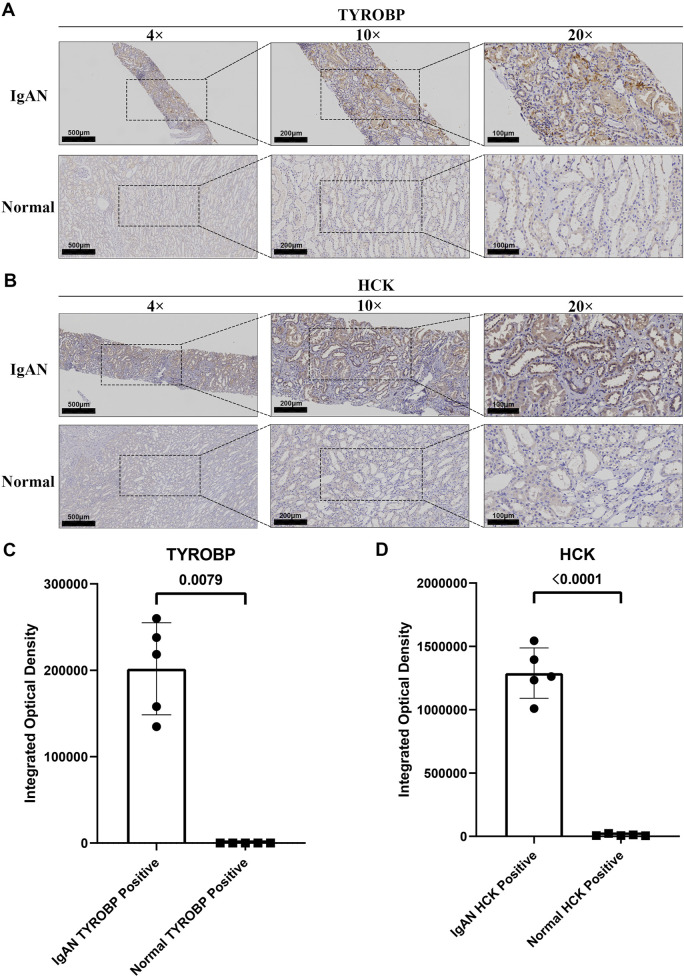
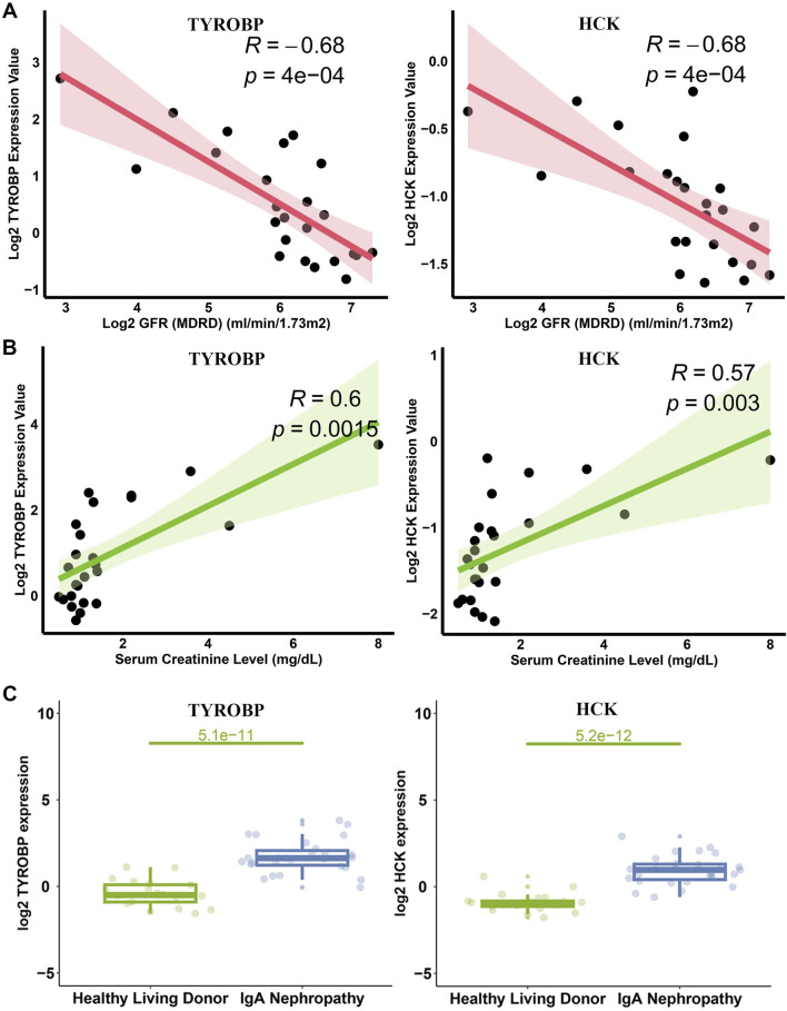
Results: Urine RNA-seq analysis revealed differential expression profiles, from which TYROBP and HCK were identified as key biomarkers using machine learning. These biomarkers were validated in both a test cohort and an external validation cohort, demonstrating strong predictive accuracy. scRNA-seq confirmed their cell-specific expression patterns, correlating with renal function metrics such as GFR and serum creatinine. IHC further validated protein expression, and molecular docking suggested potential therapeutic interactions with IgAN treatments.

Conclusion: TYROBP and HCK are promising non-invasive urinary biomarkers for IgAN. Their predictive accuracy, validated through machine learning, along with IHC confirmation and molecular docking insights, supports their potential for both diagnostic and therapeutic applications in IgAN.

Keywords: HCK; IgA nephropathy; TYROBP; molecular docking; non-invasive biomarkers; urine bulk RNA sequencing.


Conflict of interest statement

The authors declare that the research was conducted in the absence of any commercial or financial relationships that could be construed as a potential conflict of interest.

Figures

FIGURE 1
FIGURE 1
Workflow diagram.
FIGURE 2
FIGURE 2
Workflow for urine cell sampling, bulk RNA sequencing, and differential analysis (A) Workflow diagram depicting the processing of urine samples and the bulk RNA sequencing procedure. (B–D) Volcano plots illustrating differential gene expression between IgAN patients and healthy controls in first morning urine, second morning urine, and random urine samples. (E) Venn diagram showing the overlap of upregulated differential genes between IgAN patients and healthy controls across first morning urine, second morning urine, and random urine samples.
FIGURE 3
FIGURE 3
Functional insights and renal origins of commonly upregulated genes in second morning urine samples from IgAN patients (A) Differential gene analysis in IgAN kidney tissue. (B) Overlap between differential genes identified in second morning urine and those in kidney tissue. (C–F) KEGG and GO enrichment analyses highlighting associations with immune and signaling pathways.
FIGURE 4
FIGURE 4
Machine learning unveils TYROBP and HCK as core IgAN biomarkers (A) Lasso regression identifies key genes from 43 upregulated genes in second-morning urine samples. (B) Random forest analysis further refines gene selection, pinpointing pivotal biomarkers. (C) XGBoost algorithm enhances feature importance, validating key genes for IgAN. (D) Venn diagram illustrates TYROBP and HCK as core diagnostic and prognostic markers for IgAN.
FIGURE 5
FIGURE 5
Single-cell transcriptomic profiling reveals distinct cellular clusters and marker gene expression patterns (A, D) Clustering of 70,299 cells from publicly available scRNA-seq datasets (GSE131685, GSE171314, GSE140989, GSE127136) into 20 distinct clusters, categorized into 15 different cell types. (B, C) Bubble plots illustrating the expression of marker genes across the 20 clusters and 15 cell types. (E) Violin plots showing high expression of TYROBP in monocyte-macrophages and NK-T cells, and high expression of HCK specifically in monocyte-macrophages. (F) Density plots further confirming the elevated expression of TYROBP and HCK in monocyte-macrophages.
FIGURE 6
FIGURE 6
Differential expression and developmental dynamics of TYROBP and HCK in monocyte-macrophage subpopulations (A) t-SNE clustering of 3,676 monocyte-macrophage cells into four subgroups, with three of these subgroups primarily consisting of peripheral blood monocytes. (B) Violin plots showing that TYROBP is highly expressed in all subgroups, whereas HCK is predominantly expressed in subgroups 0, 1, and 3. (C) Pseudotime analysis indicates that the three subgroups predominantly composed of peripheral blood monocytes represent the developmental starting point, with cells transitioning from state 1 to state 4 along the pseudotime axis. (D) TYROBP maintains high and progressively increasing expression along the developmental trajectory, while HCK’s expression pattern inversely correlates with macrophage activation, suggesting a potential association with the activity state of monocyte-macrophages.
FIGURE 7
FIGURE 7
Elevated expression of TYROBP and HCK in IgAN renal tissues compared to normal kidney tissues. (A) Representative IHC staining of TYROBP in IgAN renal tissues and normal controls, demonstrating its upregulation in IgAN. Images are shown at 4×, ×10, and ×20 magnification. (B) Representative IHC staining of HCK in IgAN renal tissues and normal controls, revealing its increased expression in IgAN. Images are presented at 4×, ×10, and ×20 magnification. (C) Quantitative analysis of integrated optical density (IOD) for TYROBP in IgAN and normal kidney tissues. The IOD of TYROBP-positive areas in IgAN tissues is significantly higher than in normal tissues, confirming its upregulation. (D) Quantitative analysis of integrated optical density for HCK in IgAN and normal kidney tissues. The IOD of HCK-positive areas in IgAN tissues is significantly elevated compared to normal tissues, supporting its enhanced expression in IgAN.
FIGURE 8
FIGURE 8
Diagnostic efficacy of TYROBP and HCK in IgAN: ROC curves and AUC values in test and validation sets. (A, B) ROC curves for TYROBP and HCK in the test set, showing AUC values of 0.909 and 0.843, respectively, indicating strong diagnostic performance. (C) Combined ROC curve for TYROBP and HCK in the test set, achieving an AUC of 0.942, demonstrating enhanced diagnostic accuracy. (D, E) ROC curves for TYROBP and HCK in the validation sets, with AUCs of 0.906 and 0.845, respectively, confirming robustness across external datasets. (F) Combined ROC curve for TYROBP and HCK in the validation sets, with an AUC of 0.917, highlighting the biomarkers’ potential for reliable early diagnosis and clinical management of IgAN.
FIGURE 9
FIGURE 9
Correlation of TYROBP and HCK with GFR and Scr in IgAN. (A) Scatter plots depicting the inverse correlations of TYROBP and HCK with GFR in IgAN patients. (B) Scatter plots showing positive correlations of TYROBP and HCK with Scr levels. (C) Expression analysis from the Nephroseq database revealing significantly elevated levels of TYROBP and HCK in IgAN patients compared to controls.
FIGURE 10
FIGURE 10
Molecular docking analysis of diagnostic proteins TYROBP and HCK with small molecules. (A, B) Sacubitril valsartan sodium hydrate docked with TYROBP and HCK, showing strong binding affinities indicative of potential modulation of disease pathways. (C, D) Dapagliflozin interactions with TYROBP and HCK, exhibiting the lowest binding free energy among tested compounds, highlighting renal protective potential. (E, F) Budesonide docked with TYROBP and HCK, demonstrating interactions relevant to inflammation control. (G, H) Sparsentan binding to TYROBP and HCK underscores its dual receptor blockade properties, with implications for renal health management. (I, J) Bathocuproine disulfonate docked with TYROBP and HCK, emerging as a novel therapeutic candidate with promising binding energies, particularly notable for HCK.

Similar articles

References

    1. Bai Y., Li Y., Xi Y., Ma C. (2022). Identification and validation of glomerulotubular crosstalk genes mediating IgA nephropathy by integrated bioinformatics. BMC Nephrol. 23 (1), 143. 10.1186/s12882-022-02779-7 - DOI - PMC - PubMed
    1. Barratt J., Lafayette R., Kristensen J., Stone A., Cattran D., Floege J., et al. (2023b). Results from part A of the multi-center, double-blind, randomized, placebo-controlled NefIgArd trial, which evaluated targeted-release formulation of budesonide for the treatment of primary immunoglobulin A nephropathy. Kidney Int. 103 (2), 391–402. 10.1016/j.kint.2022.09.017 - DOI - PubMed
    1. Barratt J., Rovin B., Wong M. G., Alpers C. E., Bieler S., He P., et al. (2023a). IgA nephropathy patient baseline characteristics in the sparsentan PROTECT study. Kidney Int. Rep. 8 (5), 1043–1056. 10.1016/j.ekir.2023.02.1086 - DOI - PMC - PubMed
    1. Beckmann M., Wilson T., Lloyd A. J., Torres D., Goios A., Willis N. D., et al. (2020). Challenges associated with the design and deployment of food intake urine biomarker technology for assessment of habitual diet in free-living individuals and populations—a perspective. Front. Nutr. 7, 602515. 10.3389/fnut.2020.602515 - DOI - PMC - PubMed
    1. Cai G. Y., Chen X. M. (2009). Immunoglobulin A nephropathy in China: progress and challenges. Am. J. Nephrol. 30 (3), 268–273. 10.1159/000225563 - DOI - PubMed

LinkOut - more resources