. 2024 Dec 23:15:1482892.
doi: 10.3389/fneur.2024.1482892. eCollection 2024.

Myosin XVA isoforms participate in the mechanotransduction-dependent remodeling of the actin cytoskeleton in auditory stereocilia

Affiliations

Myosin XVA isoforms participate in the mechanotransduction-dependent remodeling of the actin cytoskeleton in auditory stereocilia

Ana I López-Porras et al. Front Neurol. .

Abstract

Auditory hair cells form precise and sensitive staircase-like actin protrusions known as stereocilia. These specialized microvilli detect deflections induced by sound through the activation of mechano-electrical transduction (MET) channels located at their tips. At rest, a small MET channel current results in a constant calcium influx which regulates the morphology of the actin cytoskeleton in the shorter 'transducing' stereocilia. However, the molecular mechanisms involved in this novel type of activity-driven plasticity in the stereocilium cytoskeleton are currently unknown. Here, we tested the contribution of myosin XVA (MYO15A) isoforms, given their known roles in the regulation of stereocilia dimensions during hair bundle development and the maintenance of transducing stereocilia in mature hair cells. We used electron microscopy to evaluate morphological changes in the cytoskeleton of auditory hair cell stereocilia after the pharmacological blockage of resting MET currents in cochlear explants from mice that lacked one or all isoforms of MYO15A. Hair cells lacking functional MYO15A isoforms did not exhibit MET-dependent remodeling in their stereocilia cytoskeleton. In contrast, hair cells lacking only the long isoform of MYO15A exhibited increased MET-dependent stereocilia remodeling, including remodeling in stereocilia from the tallest 'non-transducing' row of the bundle. We conclude that MYO15A isoforms both enable and fine-tune the MET-dependent remodeling of the actin cytoskeleton in transducing stereocilia, while also contributing to the stability of the tallest row.

Keywords: actin cytoskeleton; hair cells; mechanotransduction; myosin XVA; remodeling; stereocilia.


Conflict of interest statement

The authors declare that the research was conducted in the absence of any commercial or financial relationships that could be construed as a potential conflict of interest.

Figures

Figure 1
Figure 1
Outer hair cells lacking functional MYO15A do not exhibit mechanotransduction-dependent stereocilia remodeling. (A) Decreased FM1-43 dye uptake in hair cells after application of the MET channel blocker, tubocurarine (300 μM), in heterozygous (left) and homozygous shaker2 (right) cochlear explants. (B) Representative false-colored scanning electron microscopy (SEM) images of outer hair cell (OHC) stereocilia bundles cultured for 4 h at 37°C in control conditions (top) or with 300 μM tubocurarine (bottom), from heterozygous control (left) or homozygous shaker2 (right) littermates. Insets show highlighted bundle regions at higher magnification. (C) Diameter of stereocilia from the second row [colored in yellow in panel (B)] at several positions from the stereocilium tip (as indicated in cartoon), from OHC of control heterozygous (black, gray) or homozygous shaker2 (dark and light blue) littermates cultured in control conditions (darker bars) or with tubocurarine (lighter bars). Data are shown as mean ± SE. Statistical differences were analyzed using a linear mixed model. (D) Heights of stereocilia from the first, second and third rows [colored in pink, yellow and cyan in panel (B), respectively] of OHC from control heterozygous (black, gray) or homozygous shaker2 (dark and light blue) littermates cultured in control conditions (darker points) or with tubocurarine (lighter points). Horizontal lines indicate the mean and statistical differences were obtained using Welch’s t test. For all panels, the age of explants is P5, and statistical significance is shown as: *p < 0.05, **p < 0.01, ***p < 0.001, ****p < 0.0001; n.s., not significant.
Figure 2
Figure 2
Inner hair cells lacking functional MYO15A do not exhibit mechanotransduction-dependent stereocilia remodeling. (A) Representative false-colored SEM images of inner hair cell (IHC) stereocilia bundles cultured for 4 h at 37°C in control conditions (top) or with 300 μM tubocurarine (bottom), from heterozygous (left) or homozygous shaker2 (right) littermates. Insets show highlighted bundle regions at higher magnification. (B) Diameter of stereocilia from the second row [colored in yellow in panel (A)] at several positions from the stereocilium tip (as indicated in cartoon), from IHC of control heterozygous (black, gray) or homozygous shaker2 (dark and light blue) littermates cultured in control conditions (darker bars) or with tubocurarine (lighter bars). Data are shown as mean ± SE. Statistical differences were analyzed using a linear mixed model. (C) Heights of stereocilia from the first, second and third rows [colored in pink, yellow and cyan in panel (A), respectively] of IHC from control heterozygous (black, gray) or homozygous shaker2 (dark and light blue) littermates cultured in control conditions (darker points) or with tubocurarine (lighter points). Horizontal lines indicate the mean and statistical differences were obtained using Welch’s t tests. (D) Representative focused ion beam SEM (FIB-SEM) images from homozygous shaker2 mice cultured at 37°C in control conditions for 4 h (top), with 60 μM tubocurarine for 5.5 h (middle), or with 300 μM tubocurarine for 4 h (bottom). (E) Heights of stereocilia from the first and second rows of the bundle obtained from FIB-SEM images of homozygous shaker2 IHC cultured in control conditions (darker points) or with tubocurarine (lighter points). Horizontal lines indicate the mean and statistical differences were obtained using a Šídák’s multiple comparisons test. For all panels, the age of explants is P5, and statistical significance is shown as: *p < 0.05, **p < 0.01, ***p < 0.001, ****p < 0.0001; n.s., not significant.
Figure 3
Figure 3
Outer hair cells lacking the long isoform of MYO15A show accelerated mechanotransduction-dependent stereocilia remodeling. (A) Decreased FM1-43 dye uptake in hair cells after application of the MET channel blocker, tubocurarine (300 μM), in wild-type Myo15a+/+ (top), heterozygous Myo15a+/ΔN (middle) or homozygous Myo15aΔN/ΔN (bottom) littermates. (B) Representative false-colored SEM images of OHC stereocilia bundles cultured for 4 h at 37°C in control conditions (top) or with 300 μM tubocurarine (bottom), from wild-type (left), heterozygous mice (middle) or homozygous Myo15aΔN (right) littermates. Insets show highlighted bundle regions at higher magnification. (C) Diameter of stereocilia from the second row [colored in yellow in panel (B)] at several positions from the stereocilium tip from OHC of wild-type (black), heterozygous (darker shades of green) and homozygous Myo15aΔN (brighter shades of green) littermates cultured in control conditions (darker bars) or with tubocurarine (lighter bars). Data are shown as mean ± SE. Statistical differences were analyzed using a linear mixed model. (D) Stereocilia heights from the second and third rows [colored in yellow and cyan, respectively, in panel (B)] of OHC from control wild-type (black, gray), heterozygous (darker shades of green) or homozygous Myo15aΔN (brighter shades of green) littermates cultured in control conditions (darker points) or with tubocurarine (lighter points). Horizontal lines indicate the mean and statistical differences were obtained using a Šídák’s multiple comparisons test. For all panels, the age of explants is P7, and statistical significance is shown as: *p < 0.01, **p < 0.001, ***p < 0.001, ****p < 0.0001; n.s., not significant. Data are representative of four independent series with 3–4 h incubations with 300 μM tubocurarine in P4–P7 explants.
Figure 4
Figure 4
Inner hair cells lacking the long isoform of MYO15A show accelerated MET-dependent stereocilia remodeling. (A) Representative SEM images of IHC stereocilia bundles from wild-type (left), heterozygous (middle), or homozygous Myo15aΔN (right) littermates cultured for 4 h at 37°C in control conditions (top) or with 300 μM tubocurarine (bottom). Insets show highlighted bundle regions at higher magnification. (B) Diameter of stereocilia from the second row of IHC [colored in yellow in panel (A)] at several positions from the stereocilium tip, from wild-type (black), heterozygous (dark shades of green), and homozygous Myo15aΔN (bright shades of green) littermates cultured in control conditions (darker bars) or with tubocurarine (lighter bars). Data are shown as mean ± SE. Statistical differences were analyzed using a linear mixed model. (C) Stereocilia heights from the second and third row of IHC [colored in yellow and cyan, respectively, in panel (A)] from wild-type (black), heterozygous (darker shades of green), or homozygous Myo15aΔN (brighter shades of green) littermates cultured in control conditions (darker points) or with tubocurarine (lighter points). Horizontal lines indicate the mean and statistical differences were obtained using Šídák’s multiple comparisons test. For all panels, the age of explants is P7, and statistical significance is shown as: *p < 0.01, **p < 0.001, ***p < 0.001, ***p < 0.0001; n.s., not significant. Data are representative of four independent series with 3–4 h incubations with 300 μM tubocurarine in P4–P7 explants.
Figure 5
Figure 5
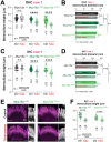
Hair cells lacking the long isoform of MYO15A exhibit MET-dependent stereocilia remodeling in the tallest row of the bundle. (A–D) Stereocilia heights (A,C) and diameters (B,D) from the first row of OHC (A,B) and IHC (C,D) from wild-type (black and gray), heterozygous (darker shades of green) or homozygous Myo15aΔN (brighter shades of green) littermates that were cultured in control conditions (darker points/bars) or with 300 μM tubocurarine (lighter points/bars) for 4 h at 37°C. Data are representative of four independent series with 3–4 h incubations with 300 μM tubocurarine in P4–P7 explants. (E) Confocal images of IHC stereocilia stained against F-actin with fluorescently-labeled phalloidin (magenta), cultured for 4 h at 37°C in control conditions (top) or with 300 μM tubocurarine (bottom), from heterozygous (left) and homozygous Myo15aΔN (right) littermates. Insets show bundle regions with Imaris volume render at higher magnification. Images are representative of two independent experiments. (F) Stereocilia heights from the first row of IHC from heterozygous (dark shades of green) or homozygous Myo15aΔN (bright shades of green) littermates cultured in control conditions (darker points) or with tubocurarine (lighter points). Stereocilia heights were measured from Imaris volume rendering of the confocal stacks. For all panels, the age of explants is P7, and statistical significance is shown as: *p < 0.01, **p < 0.001, ***p < 0.001, ****p < 0.0001; n.s., not significant. In panels (B) and (D), significant differences are shown for control conditions between genotypes (black, relative to wild-type), or between control and tubocurarine conditions within each genotype (color coded for each genotype: black for wild-type, dark green for heterozygous, and bright green for homozygous Myo15aΔN littermates).
Figure 6
Figure 6
Impaired expression of ESPNL in inner hair cells lacking the long isoform of MYO15A. Maximum intensity projections of confocal stacks of IHC stereocilia from the apical (A) or middle (B) cochlear regions, at postnatal days 3 (A) or 7 (B), from wild-type (left), heterozygous (middle), and homozygous Myo15aΔN (right) mice immunolabeled against ESPNL (red) and counterstained against F-actin with fluorescently-labeled phalloidin (blue). Images are representative of two independent experiments, with similar labeling patterns along different cochlear regions.
Figure 7
Figure 7
GNAI3 is mislocalized in outer, but not inner, hair cells lacking the long isoform of MYO15A. Maximum intensity projections of confocal stacks of OHC (A,C) and IHC (B,D) stereocilia from wild-type (left), heterozygous (middle), and homozygous Myo15aΔN (right) mice immunolabeled against EPS8 (green) (A,B) or GNAI3 (C,D), and counterstained against F-actin with fluorescently-labeled phalloidin (red). Yellow arrowheads point to OHC regions lacking GNAI3 labeling in heterozygous and homozygous Myo15aΔN mice. In panels (C) and (D), the maximum intensity projections ignored Z planes near the cuticular plate to avoid confounding GNAI3 labeling from the bare zone of hair cells. Images are representative of two independent experiments. To see the GNAI3 labeling in the bare zone, see Supplementary Figure 8. In all panels the age of explants is P7.

Update of

Similar articles

Cited by

References

    1. Shin JB, Longo-Guess CM, Gagnon LH, Saylor KW, Dumont RA, Spinelli KJ, et al. . The R109H variant of fascin-2, a developmentally regulated actin crosslinker in hair-cell stereocilia, underlies early-onset hearing loss of dba/2J mice. J Neurosci. (2010) 30:9683–94. doi: 10.1523/JNEUROSCI.1541-10.2010, PMID: - DOI - PMC - PubMed
    1. Taylor R, Bullen A, Johnson SL, Grimm-Gunter EM, Rivero F, Marcotti W, et al. . Absence of plastin 1 causes abnormal maintenance of hair cell stereocilia and a moderate form of hearing loss in mice. Hum Mol Genet. (2015) 24:37–49. doi: 10.1093/hmg/ddu417, PMID: - DOI - PMC - PubMed
    1. Tilney LG, Derosier DJ, Mulroy MJ. The organization of actin filaments in the stereocilia of cochlear hair cells. J Cell Biol. (1980) 86:244–59. doi: 10.1083/jcb.86.1.244, PMID: - DOI - PMC - PubMed
    1. Zheng L, Sekerkova G, Vranich K, Tilney LG, Mugnaini E, Bartles JR. The deaf jerker mouse has a mutation in the gene encoding the espin actin-bundling proteins of hair cell stereocilia and lacks espins. Cell. (2000) 102:377–85. doi: 10.1016/S0092-8674(00)00042-8, PMID: - DOI - PMC - PubMed
    1. Assad JA, Shepherd GM, Corey DP. Tip-link integrity and mechanical transduction in vertebrate hair cells. Neuron. (1991) 7:985–94. doi: 10.1016/0896-6273(91)90343-X, PMID: - DOI - PubMed

LinkOut - more resources