Review
. 2024 Nov 15:89:e531-e540.
doi: 10.5114/pjr/192424. eCollection 2024.

Imaging of intramedullary tumours of the spinal cord

Affiliations
Review

Imaging of intramedullary tumours of the spinal cord

Marek Sąsiadek et al. Pol J Radiol. .

Abstract

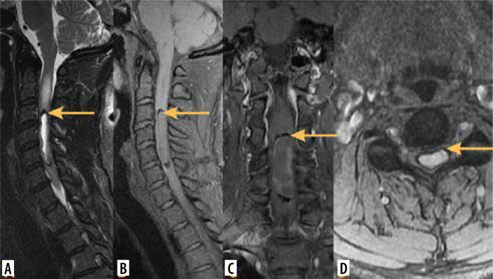
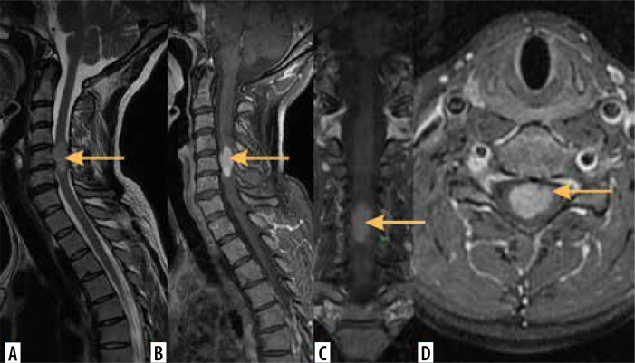
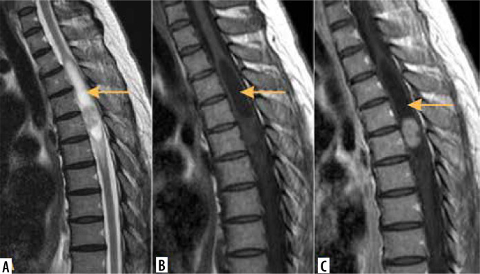
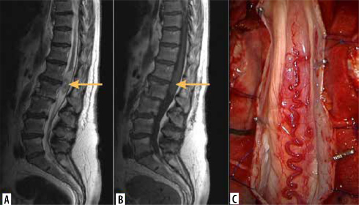
Intramedullary tumours (IMTs) are the least common neoplasms of the spinal canal. The majority of them are ependymomas and astrocytomas, the third commonest is haemangioblastoma, while other tumours of the spinal cord are relatively rare. This review presents on update on the imaging of spinal cord tumours. Magnetic resonance imaging (MRI) is the imaging method of choice in diagnosing IMTs, with other modalities playing a supplementary role. The authors discuss the MRI protocol in IMTs including advanced techniques and present the imaging features of particular tumours. The differentiation of IMTs from other spinal cord diseases is also presented.

Keywords: MRI; intramedullary tumours; spine imaging.


Figures

Figure 1
Figure 1
Ependymoma. Male, 45 years. MR sagittal T2-weighted (A), fat suppression sagittal (B), coronal (C) and axial (D) images. Note the typical peripheral rim of hemosiderin (arrows)
Figure 2
Figure 2
Ependymoma. Female, 45 years with progressive paraparesis. MR sagittal T2-weighted (A), T1-weighted post-gadolinium sagittal (B), coronal (C) and axial (D) images. Note the central location of the tumour in the spinal cord (arrows)
Figure 3
Figure 3
Ependymoma, WHO, grade 2. Female, 41 years. MR sagittal T2-weighted (A), T1-weighted (B) and T1-weighted post-gadolinium (C) images. Note the syringomyelic cavity above the tumour (arrows)
Figure 4
Figure 4
Anaplastic ependymoma, WHO, grade 3. MR sagittal T2-weighted (A), and T1-weighted (B) images. Tumour is located in the conus medullaris (arrows). C) Intraoperative photo of the tumour (courtesy of Mr. Marcel Ivanov, MD, Consultant Spinal Neurosurgeon, Sheffield Teaching Hospitals NHS Foundation Trust, Sheffield, UK)
Figure 5
Figure 5
Neurofibromatosis type 2. MR sagittal T2-weighted (A, B), and T1-weighted post-gadolinium (C, D) images. Intramedullary tumour (ependymoma) in the cervical spinal cord (arrows). Multiple schwannomas/meningiomas in the intradural extramedullary space (arrowheads)
Figure 6
Figure 6
Myxopapillary ependymoma. MR sagittal T2-weighted (A, B), T1-weighted (C) and T1-weighted post-gadolinium (D) images. Tumour located in the conus medullaris/filum terminale region (arrows)
Figure 7
Figure 7
Astrocytoma. Male, 6 years with progressive paraparesis. MR sagittal T2-weighted (A), T1-weighted (B), post-gadolinium T1-weighted sagittal (C) and coronal (D) images. Note the eccentric location of the tumour, marked widening of the spinal cord, irregular contrast enhancement as well as multiple intratumoural cysts (arrow on A)
Figure 8
Figure 8
Pilocytic astrocytoma, WHO, grade 1 in the thoracic spinal cord. MR axial T2-weighted (A), axial T1-weighted post-gadolinium (B), sagittal T1-weighted (C), sagittal T1-weighted post-gadolinium (D) images (arrows). (E) Intraoperative photo of the tumour (courtesy of Mr. Marcel Ivanov, MD, Consultant Spinal Neurosurgeon, Sheffield Teaching Hospitals NHS Foundation Trust, Sheffield, UK)
Figure 9
Figure 9
Astrocytoma in upper cervical spinal cord. Male, 20 years. MR sagittal T2-weighted (A, B), coronal T2-weighted (C) and post-gadolinium T1-weighted sagittal (D) images. Note the eccentrical location of the tumour, widening of the spinal cord and small intratumoural cysts (arrows)
Figure 10
Figure 10
Astrocytoma in thoracic spinal cord. MR sagittal T2-weighted (A), coronal (B) and sagittal (C) diffusion tensor tractography (DTT) images. Note the smooth margins of the tumour in DTT images (arrows), which suggest its expansion without infiltration of the adjacent spinal cord tissue
Figure 11
Figure 11
Haemangioblastoma. MR sagittal T2-weighted (A), T1-weighted (B) and T1-weighted post-gadolinium (C), axial T2-weighted (D) and T1-weighted post-gadolinium (E) images. Note large cystic component (arrows) and small enhancing marginal nodule (arrowhead)
Figure 12
Figure 12
Paraganglioma of filum terminale. MR sagittal T2-weighted images (A, B). There are multiple linear signal void foci compatible with abnormal vessels both within and above the tumour (arrowheads)
Figure 13
Figure 13
Dermoid cyst of conus medullaris. MR sagittal T2-weighted (A) and T1-weighted (non-enhanced) (B) images. Note high signal focus on T1-weighted image, compatible with fatty component (arrow on B)
Figure 14
Figure 14
Differentiation: multiple sclerosis. MR sagittal (A) and coronal (B) T2-weighted images. Multiple hyperintense foci in the spinal cord (arrows)
Figure 15
Figure 15
Differentiation: syringomyelia. MR sagittal T2-weighted (A), T1-weighted (B) and T1-weighted post-gadolinium sagittal (C) and axial (D) images. Note syringomyelic cavity without contrast enhancement (arrows on C and D)
Figure 16
Figure 16
Differentiation: spinal dural arterio-venous fistula (SDAVF). MR axial (A) and sagittal (B) T2-weighted images. Diffuse enlargement and hyperintensity of the spinal cord with multiple tiny linear signal void foci on the posterior surface of the spinal cord (arrows), compatible with abnormal vessels. (C) Spinal angiography confirms diagnosis of SDAVF

Similar articles

References

    1. Das JM, Hoang S, Mesfin FB. Intramedullary spinal cord tumors. In: StatPearls; Treasure Island, FL: StatPearls Publishing; 2023.
    1. Neal MT, Richards AE, Curley KL, Donev K, Lyons MK, Kalani MA. Spinal intramedullary hemangioblastoma and schwannoma collision tumor: illustrative case. J Neurosurg Case Lessons 2021; 1: CASE2059. DOI: 10.3171/CASE2059. - DOI - PMC - PubMed
    1. Merhemic Z, Stosic-Opincal T, Thurnher MM. Neuroimaging of spinal tumors. Magn Reson Imaging Clin N Am 2016; 24: 563-579. - PubMed
    1. Pinter NK, Pfiffner TJ, Mechtler LL. Neuroimaging of spine tumors. Handb Clin Neurol 2016; 136: 689-706. - PubMed
    1. Shah LM, Salzman KL. Conventional and advanced imaging of spinal cord tumors. Neuroimaging Clin N Am 2023; 33: 389-406. - PubMed

LinkOut - more resources