. 2025 Jan 8;23(1):e3002977.
doi: 10.1371/journal.pbio.3002977. eCollection 2025 Jan.

Are oligodendrocytes bystanders or drivers of Parkinson's disease pathology?

Affiliations

Are oligodendrocytes bystanders or drivers of Parkinson's disease pathology?

José María Salazar Campos et al. PLoS Biol. .

Abstract

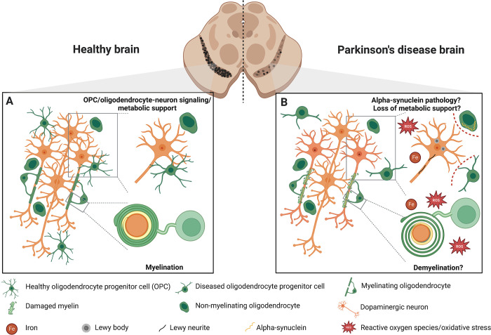
The major pathological feature of Parkinson 's disease (PD), the second most common neurodegenerative disease and most common movement disorder, is the predominant degeneration of dopaminergic neurons in the substantia nigra, a part of the midbrain. Despite decades of research, the molecular mechanisms of the origin of the disease remain unknown. While the disease was initially viewed as a purely neuronal disorder, results from single-cell transcriptomics have suggested that oligodendrocytes may play an important role in the early stages of Parkinson's. Although these findings are of high relevance, particularly to the search for effective disease-modifying therapies, the actual functional role of oligodendrocytes in Parkinson's disease remains highly speculative and requires a concerted scientific effort to be better understood. This Unsolved Mystery discusses the limited understanding of oligodendrocytes in PD, highlighting unresolved questions regarding functional changes in oligodendroglia, the role of myelin in nigral dopaminergic neurons, the impact of the toxic environment, and the aggregation of alpha-synuclein within oligodendrocytes.


Conflict of interest statement

The authors have declared that no competing interests exist.

Figures

Fig 1
Fig 1. Disease modeling in PD.
PD is a complex neurodegenerative human pathology that on the tissue level (A) is characterized by the loss of dopaminergic neurons in the substantia nigra in the human midbrain. On the cellular level (B), the major hallmark of PD pathology is the intracellular accumulation of α-synuclein into Lewy bodies and Lewy neurites in dopaminergic neurons and dopaminergic neuron degeneration. To model PD pathology, many genetic and toxin-induced mouse models (reviewed in [5]) have been developed (C). However, neither of those fully recapitulates human pathology, which is also likely due to species-specific differences between rodent and human biology [6]. Only a few models have specifically examined oligodendrocytes [7]. Therefore, human iPSC-derived 2D [8] and 3D models have been developed, including models with oligodendrocytes [9] with increasingly complex cellular compositions (D). Created with BioRender.com. iPSC, induced pluripotent stem cell; PD, Parkinson’s disease.
Fig 2
Fig 2. Function and dysfunction of oligodendrocyte lineage cells in healthy and PD brains.
(A) In the healthy brain, the primary role of oligodendrocytes is the myelination of axons, though dopaminergic neurons are generally thought to have minimal myelination [14]. Moreover, both OPCs and non-myelinating oligodendrocytes are recognized for their crucial roles in signaling and providing metabolic support [10], although the exact function of the non-myelinating oligodendrocytes is even less clear. (B) In the PD brain, the particular consequences of damaged or non-functional oligodendrocytes is not well understood. Although there are reports of demyelination in patient brains [12,13], it is still unclear to what extent this affects dopaminergic neuron health or whether demyelination precedes neuronal damage. α-synuclein positive inclusions have been found in oligodendrocytes in PD patient brains [29,30]; however, it remains unclear whether this is due to cell-autonomous build-up of α-synuclein or protein cell-to-cell transfer from neurons to oligodendrocytes. It is hypothesized that diseased oligodendrocytes and OPCs may lose their direct signaling and metabolic support functions (indicated by dashed red lines) to neurons due to pathological changes in their environment, such as the accumulation of ROS and iron (Fe) accumulation that could exacerbate neuronal damage. Created with BioRender.com. OPC, oligodendrocyte progenitor cell; PD, Parkinson’s disease; ROS, reactive oxygen species.

Similar articles

References

    1. Spillantini MG, Schmidt ML, Lee VM, Trojanowski JQ, Jakes R, Goedert M. Alpha-synuclein in Lewy bodies. Nature. 1997;388(6645):839–40. doi: 10.1038/42166 . - DOI - PubMed
    1. Wong YC, Krainc D. α-synuclein toxicity in neurodegeneration: mechanism and therapeutic strategies. Nat Med. 2017;23(2):1–13. - PMC - PubMed
    1. Kamath T, Abdulraouf A, Burris SJ, Langlieb J, Gazestani V, Nadaf NM, et al.. Single-cell genomic profiling of human dopamine neurons identifies a population that selectively degenerates in Parkinson’s disease. Nat Neurosci. 2022;25(5):588–95. doi: 10.1038/s41593-022-01061-1 - DOI - PMC - PubMed
    1. Okano H, Morimoto S. iPSC-based disease modeling and drug discovery in cardinal neurodegenerative disorders. Cell Stem Cell. 2022;29(2):189–208. doi: 10.1016/j.stem.2022.01.007 . - DOI - PubMed
    1. Chia SJ, Tan E-K, Chao Y-X. Historical Perspective: Models of Parkinson’s Disease. Int J Mol Sci. 2020;21(7). doi: 10.3390/ijms21072464 ; PubMed Central PMCID: PMC7177377. - DOI - PMC - PubMed

MeSH terms

Substances