. 2025 Jan 7;53(1):gkae1250.
doi: 10.1093/nar/gkae1250.

Design and in vitro anticancer assessment of a click chemistry-derived dinuclear copper artificial metallo-nuclease

Affiliations

Design and in vitro anticancer assessment of a click chemistry-derived dinuclear copper artificial metallo-nuclease

Simon Poole et al. Nucleic Acids Res. .

Abstract

Copper compounds with artificial metallo-nuclease (AMN) activity are mechanistically unique compared to established metallodrugs. Here, we describe the development of a new dinuclear copper AMN, Cu2-BPL-C6 (BPL-C6 = bis-1,10-phenanthroline-carbon-6), prepared using click chemistry that demonstrates site-specific DNA recognition with low micromolar cleavage activity. The BPL-C6 ligand was designed to force two redox-active copper centres-central for enhancing AMN activity-to bind DNA, via two phenanthroline ligands separated by an aliphatic linker. DNA-binding experiments, involving circular dichroism spectroscopy, agarose gel electrophoresis and fluorescence quenching, revealed a preference for binding with adenine-thymine-rich DNA. The oxidative cleavage mechanism of Cu2-BPL-C6 was then elucidated using in vitro molecular and biophysical assays, including in-liquid atomic force microscopy analysis, revealing potent DNA cleavage mediated via superoxide and hydrogen peroxide oxidative pathways. Single-molecule analysis with peripheral blood mononuclear cells identified upregulated single-strand DNA lesions in Cu2-BPL-C6-treated cells. Using specific base excision repair (BER) enzymes, we showed that Endo IV selectively repairs these lesions indicating that the complex generates apurinic and apyrimidinic adducts. Broad spectrum anticancer evaluation of BPL-C6 was performed by the National Cancer Institute's 60 human cell line screen (NCI-60) and revealed selectivity for certain melanoma, breast, colon and non-small cell lung cancer cell lines.


Figures

Graphical Abstract
Graphical Abstract
Figure 1.
Figure 1.
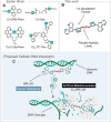
(A) The structures of earlier reported AMNs Cu-2-Clip-Phen, Cu-DPQ-Phen, Cu-Oda and Cu3-TC-Thio. (B) Molecular structure of BPL-C6 prepared using CuAAC click chemistry. (C) Upon coordination of two copper ions, Cu2-BPL-C6 promotes anticancer activity mediated by AMN activity on genomic DNA.
Figure 2.
Figure 2.
Synthetic route for the generation of Cu2-BPL-C6.
Figure 3.
Figure 3.
(A) EtBr displacement assay with BPL-C6 in the presence of titrated copper(II). (B) Binding profile of Cu2-BPL-C6 with poly[d(A-T)2] and poly[d(G-C)2], determined by the displacement of EtBr. (C) Quenching of limited bound EtBr bound to synthetic copolymers of poly[d(A-T)2] and poly[d(G-C)2] by Cu2-BPL-C6. (D) ctDNA viscosity profile of Cu2-BPL-C6 along with EtBr and Cu2TPNap controls (29,43). (E) C50 values for EtBr fluorescence quenching upon titration of Cu2-BPL-C6 2:1; Apparent binding constants of Cu2-BPL-C6 with poly[d(A-T)2], ctDNA and poly[d(G-C)2]; Q-value (EC50) of Cu2-BPL-C6 with ctDNA poly[d(A-T)2] and poly[d(G-C)2] (EtBr limited-bound).
Figure 4.
Figure 4.
Cu2-BPL-C6 interaction with ctDNA (left), poly[d(A-T)2] (middle) and poly[d(G-C)2] (right) monitored using CD spectroscopy between r = 0 and r = 0.2, where r = [Drug]/[DNA].
Figure 5.
Figure 5.
Cu2-BPL-C6 interaction with FRET labelled DNA hairpins (A) FRET-1 (B) FRET-2 (C) FRET-3 and (D) FRET-4 with increasing concentrations of complex. Bard analysis of FRET labelled DNA hairpins (E) FRET-2 and (F) FRET-1 at increasing concentrations of Cu2-BPL-C6.
Figure 6.
Figure 6.
Normalized initial fluorescence data obtained from MST experiments with (A) F-1, (B) F-2 (C) F-3 and (D) F-4, all labelled with 5′-Alexafluor647. (E) Overlay of DNA hairpins analysed with MST. (F) Table indicating the EC50 values obtained [M] for the interaction of Cu2-BPL-C6 with each DNA hairpin.
Figure 7.
Figure 7.
(A) Cleavage profile for supercoiled pUC19 DNA in the presence of Cu2-BPL-C6. (B) DNA cleavage profile of Cu2-BPL-C6 in the presence of scavengers DMTU, NaN3, tiron and D-mannitol, L-methionine and L-histidine. At the highest tested complex concentration in the presence of tiron, all three forms of pUC19 are present (lane 30) and DNA condensation is observed. In comparison, the control lane at the same concentration (lane 8) has no observable SC DNA. (C) Topoisomerase I inhibition by Cu2-BPL-C6, displaying negatively supercoiled DNA (lanes 37–43) and positively supercoiled DNA (lanes 45–49) together with evidence of DNA nicking.
Figure 8.
Figure 8.
(A) Composite image of the self-activation profile of Cu2-BPL-C6 with supercoiled pUC19 DNA in the absence of reductant at 30, 60 and 180 min time points. (B) Self-activation ROS scavenger profile of Cu2-BPL-C6 in the absence of reductant with NaN3 and D-Mannitol, tiron, DMTU, L-methionine and L-histidine after a 60 min incubation.
Figure 9.
Figure 9.
In-liquid AFM showing the self-activation activity of 20 μM Cu2-BPL-C6 on pUC19. (A) Representative AFM images of untreated pUC19, Cu2-BPL-C6 treated pUC19 after 0, 30, 60 and 180 min and the 180 min time point after the addition of 200 μM EDTA. Scale bars = 200 nm, height scales = −3 to 4 nm. (B) Quantification of the proportion of circular and linear molecules present in the images. (C) Quantitative analysis of the smallest bounding area of individual circular molecules. (D) The total volume of the masked grains. N-values are as follows: pUC19 only: 213; 0 min: 110, 30 min: 140, 60 min: 127, 180 min: 69, 180 min + EDTA: 79.
Figure 10.
Figure 10.
DNA damaging effect of Cu2-BPL-C6 on PBMCs. (A) Sample collection and treatment with Cu2-BPL-C6. (B) DNA repair with fluorescently labelled bases and post staining with YOYO-1 dye. (C) Microscopic images of control (untreated) DNA (top) and Cu2-BPL-C6 (150 μM) treated DNA (bottom) isolated from PBMCs (scale bar = 10 μm). (D) DNA damage detection in the presence of a repair enzyme cocktail for PBMCs treated with Cu2-BPL-C6 with and without antioxidant scavengers. (E) Identification of lesions generated by Cu2-BPL-C6 treated with and without BER enzymes, along with a combination thereof (cocktail).
Figure 11.
Figure 11.
(A) NCI-60 panel results indicating the lethality of BPL-C6 with non-small cell lung cancers, colon cancers, melanoma and breast cancers. (B) Table of cell lines BPL-C6 shows activity toward. (C) COMPARE analysis of BPL-C6 with standard agents available on the NCI-60 COMPARE database, indicating their Pearson correlation using LC50 data. (Signalling agents, DNA damaging agents, tubulin-directed agents, anthracyclines/topoisomerase poisons, antimetabolites/nucleosides, hormonal agents, Cu(II) agents and others.)
Figure 12.
Figure 12.
(A) Cellular viability assessment of BPL-C6 and Cu2-BPL-C6 with MDA-MB-468; (B) MDA-MB-231; (C) BT-549; (D) Table indicating IC50 data of BPL-C6 and Cu2-BPL-C6 with select cell lines.

References

    1. Watson J.D., Crick F.H.C. Molecular structure of nucleic acids: a structure for deoxyribose nucleic acid. Nature. 1953; 171:737–738. - PubMed
    1. Sigman D.S., Mazumder A., Perrin D.M. Chemical nucleases. Chem. Rev. 1993; 93:2295–2316.
    1. Sigman D.S., Graham D.R., D’Aurora V., Stern A.M. Oxygen-dependent cleavage of DNA by the 1,10-phenanthroline. cuprous complex. Inhibition of Escherichia coli DNA polymerase I. J. Biol. Chem. 1979; 254:12269–12272. - PubMed
    1. Li F., Xie J., Feng F. Copper and zinc complexes of a diaza-crown ether as artificial nucleases for the efficient hydrolytic cleavage of DNA. New J. Chem. 2015; 39:5654–5660.
    1. Zehra S., Tabassum S., Al-Lohedan H.A., Arjmand F. A zwitterionic Zn(II) benzothiazole nanohybrid conjugate as hydrolytic DNA cleavage agent. Inorg. Chem. Commun. 2018; 93:69–72.

LinkOut - more resources