. 2025 Jan 23;68(2):1179-1194.
doi: 10.1021/acs.jmedchem.4c01368. Epub 2025 Jan 8.

Modulation of Mitochondria-Endoplasmic Reticulum Contacts (MERCs) by Small Molecules as a New Strategy for Restoring Lipid Metabolism in an Amyotrophic Lateral Sclerosis Model

Affiliations

Modulation of Mitochondria-Endoplasmic Reticulum Contacts (MERCs) by Small Molecules as a New Strategy for Restoring Lipid Metabolism in an Amyotrophic Lateral Sclerosis Model

Mikel Etxebeste-Mitxeltorena et al. J Med Chem. .

Abstract

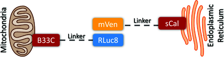
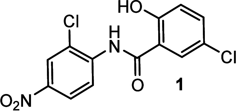
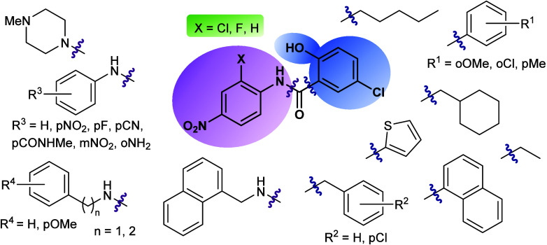
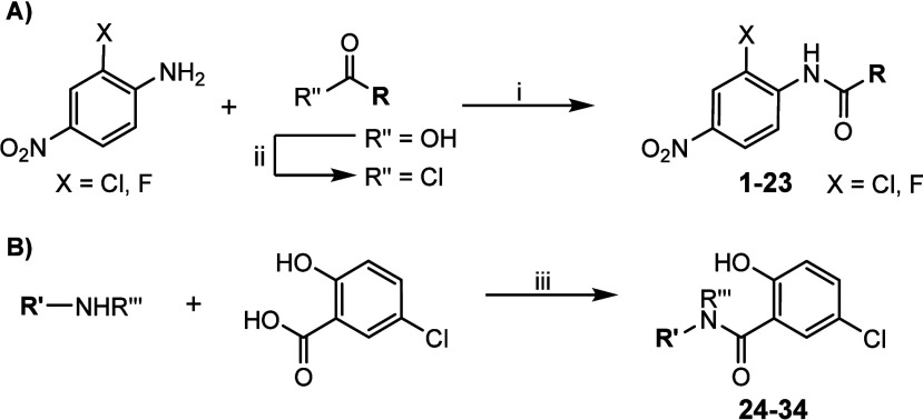
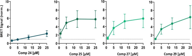
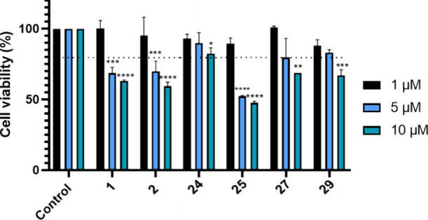
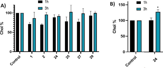
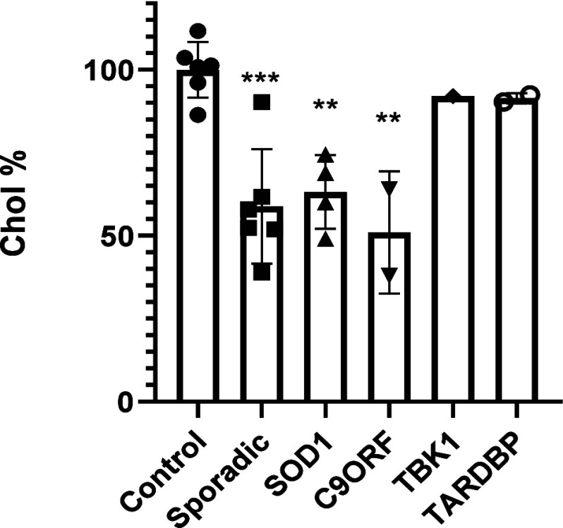
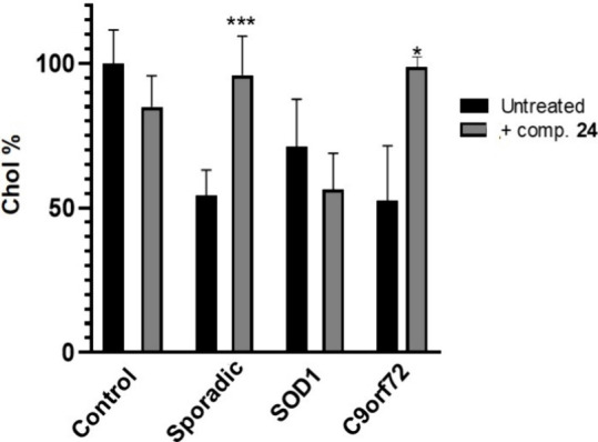
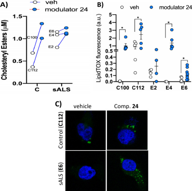
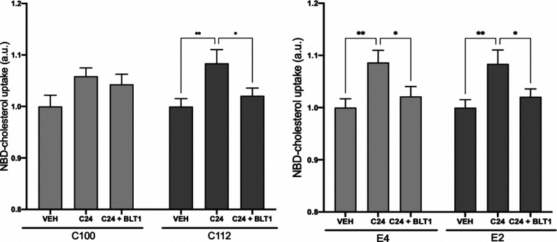
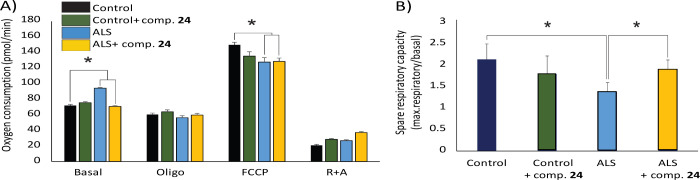
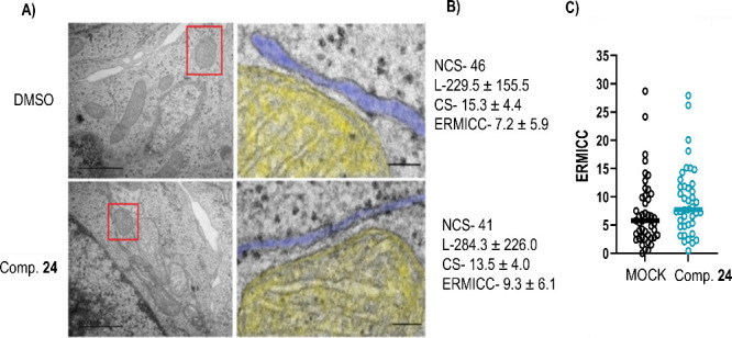
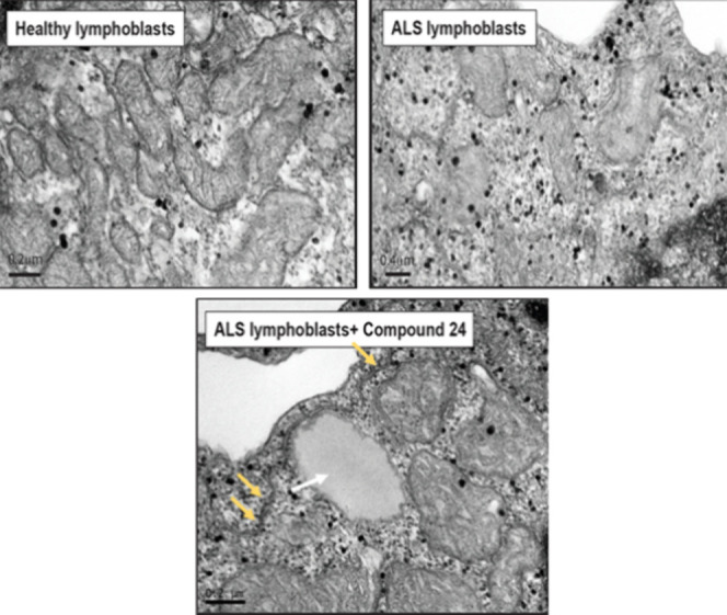
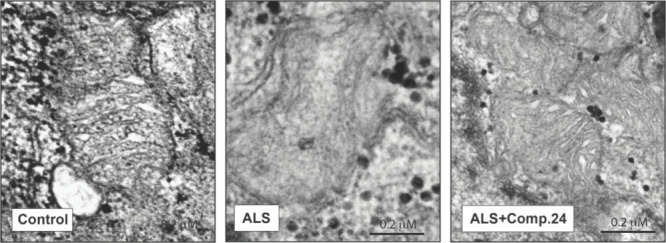
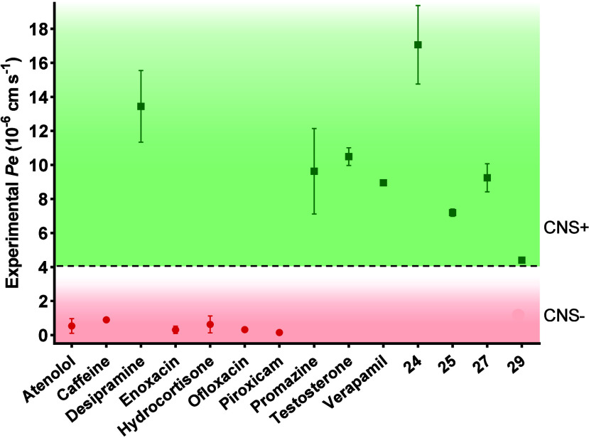
Amyotrophic lateral sclerosis (ALS) is a fatal neurodegenerative disease without effective treatment. The progressive motoneuron death in ALS is associated with alterations in lipid metabolism. As its regulation occurs in mitochondria-associated endoplasmic reticulum (ER) membranes (MAMs), modulation of mitochondria-ER contacts (MERCs) is emerging as a crucial factor in MAM formation and lipid metabolism control. Using the MERLIN biosensor in a high-throughput screening within the EU-OPENSCREEN ERIC, we discovered small molecules that increase MERCs in HCT116 cells, enhancing their ability to uptake cholesterol. We demonstrated that cholesterol trafficking is decreased in an ALS patient-derived cell model, and this trafficking is restored after treatment with the discovered MERC modulator 24. Electron microscopy revealed that treatment with compound 24 increases MERCs, promotes lipid droplet formation, and restores mitochondrial cristae. Overall, the brain-permeable MERC modulator, compound 24, may serve as a valuable pharmacological tool for studying MAM function and holds potential for in vivo studies in ALS and other MAM dysfunction diseases.


Conflict of interest statement

The authors declare no competing financial interest.

Figures

Figure 1
Figure 1
Scheme illustrating the structure of the MERLIN BRET biosensor. In MERLIN BRET pair is formed by Renilla luciferase 8 (Rluc8) targeted to the mitochondria with the C-terminal of BCL-XL (B33C), acting as donor, and mVenus (mVen) targeted to the ER by the N-terminal of S-calnexin (sCal), performing as acceptor. When in close proximity and upon the addition of cell permeable coelenterazine h, Rluc8 processes coelenterazine h into coelenteramide, excited product that can transfer the energy to the acceptor by resonance.
Figure 2
Figure 2
Chemical structure of niclosamide (1).
Figure 3
Figure 3
Chemical-related niclosamide derivatives prepared and evaluated as MERCs modulators.
Scheme 1
Scheme 1. Synthesis of the Two Series of Niclosamide Derivatives: (A) 2-Chloro- or 2-Fluoro-4-nitrophenylamide Series and (B) 5-Chloro-2-hydroxybenzamide Series
Reagents and conditions: (i) MW, 150 °C, 10 min; (ii) SOCl2, 80 °C, 2 h; (iii) EDC, HOBt, Et3N, CHCl3, 12 h.
Figure 4
Figure 4
Quantification of BRET signal alterations upon the incubation with individual MERCs modulators in HCT116 cells containing MERLIN. BRET signal, inversely correlated with mitochondria-ER distance, was measured upon the addition of compounds 24, 25, 27, and 29 at different concentrations and normalized to DMSO (MOCK) treated cells (dashed lines). Data represent the mean ± SD, N ≥ 3 independent experiments with three technical replicates per condition.
Figure 5
Figure 5
Cell viability (HCT116 cell lines) using MTT assay after treatment with compounds 1, 2, 24, 25, 27, and 29 at 1, 5, and 10 μM doses, for 24 h. Results are expressed as the mean ± standard error of the mean (SEM) for three independent experiments. *, p < 0.1; **, p < 0.01; ***, p < 0.001; ****, p < 0.0001.
Figure 6
Figure 6
Extracellular cholesterol uptake capacity of HCT166 cells after treatment with (A) compounds 1, 2, 24, 25, 27, and 29 at 1 μM for 1 and 3 h; (B) with compound 24 at 5 μM for 1 and 3 h. Results are expressed as the mean ± standard error of the mean (SEM) for three independent experiments. *, p < 0.1.
Figure 7
Figure 7
Extracellular cholesterol uptake capacity of the different familial and sporadic ALS patient’s lymphoblastic cell lines. Results are expressed as the mean ± standard error of the mean (SEM) for three independent experiments. **, p < 0.01; ***, p < 0.001.
Figure 8
Figure 8
Extracellular cholesterol uptake capacity of the different patient lymphoblastic cell lines without treatment and after treatment with compound 24 (5 μM, 24 h). Results are expressed as the mean ± standard error of the mean (SEM) for three independent experiments. *, p < 0.1; ***, p < 0.001.
Figure 9
Figure 9
Cholesteryl esters (A) and lipid droplets (B,C) were quantified in control and sALS lymphoblasts treated with MERC modulator 24 (5 μM for 24 h) or vehicle. Individual values are shown for cholesteryl esters quantification. Lipid droplet staining results are expressed as the mean ± standard error of the mean (SEM). More than 100 cells were quantified per condition. *, p < 0.1. Representative images are included (C). Nuclei were stained using DAPI (in blue). Scale bar = 20 μm.
Figure 10
Figure 10
Extracellular cholesterol uptake capacity of the different control (C100 and C112) and sALS (E2 and E4) patient lymphoblastic cell lines without treatment and after treatment with compound 24 (5 μM, 24 h). Additionally, cells were treated with 10 μM BLT1 before addition of the fluorescent NBD-cholesterol. Results are expressed as the mean ± standard error of the mean (SEM) for three independent experiments. *, p < 0.1; **, p < 0.01.
Figure 11
Figure 11
Mitochondrial oxygen consumption rates (OCR) and spare respiratory capacity in lymphoblasts derived from a healthy control and one sALS patient with and without treatment with MERCs modulator 24. (A) Basal OCR, and after addition of oligomycin, FCCP and rotenone + antimycin. (B) Spare respiratory capacity, calculated as the difference between maximal and baseline OCR. The data show the mean of four experimental replicates ± the standard error of the mean.
Figure 12
Figure 12
Effect of compound 24 on MERCs formation in HCT116 cells. (A) Left, Representative TEM-images of a mitochondria-ER contact site in HCT116 cells treated with 24 or with DMSO (MOCK). Right, zoomed area (marked with a red box). Mitochondria is shown in yellow; the ER appears in blue. Scale bar, 100 nm. (B) Quantification of MERCs on compound 24 treated and untreated cells. NCS- Number of contact sites in N = 5 cells. L- Average Length of the contact sites measured in nm. CS-Contact site, minimal distance between organelles in nm. ERMICC- ER-Mitochondria contact coefficient. (C) ERMICC coefficient of individual contact sites in DMSO and compound 24 treated cells.
Figure 13
Figure 13
TEM images on control, sALS and sALS with MERCs modulator 24 (5 μM for 24 h). The white arrow shows a lipid droplet and the yellow ones remark the MERCs.
Figure 14
Figure 14
TEM images of control, ALS and ALS + compound 24. In ALS lymphoblast disorganized or absent cristae inside the mitochondria is observed in comparison to healthy controls lymphoblasts. Treatment with compound 24 (5 μM for 24 h) restored mitochondria structure.
Figure 15
Figure 15
Permeability (Pe 10–6 cm s–1) across the BBB of control drugs (used in the experiment validation) and MERCs modulators 24, 25, 27, and 29 evaluated by the PAMPA methodology (CNS+, permeable, green; CNS–, not permeable, red).

Similar articles

Cited by

References

    1. Talbott E. O.; Malek A. M.; Lacomis D.. The epidemiology of amyotrophic lateral sclerosis. In Neuroepidemiology; Rosano C.; Ikram M. A.; Ganguli M., Eds.; Elsevier: Amsterdam, The Netherlands, 2016; Vol. 138, pp 225–238. - PubMed
    1. Ilieva H.; Vullaganti M.; Kwan J. Advances in molecular pathology, diagnosis, and treatment of amyotrophic lateral sclerosis. BMJ. 2023, 383, e07503710.1136/bmj-2023-075037. - DOI - PMC - PubMed
    1. Genin E. C.; Abou-Ali M.; Paquis-Flucklinger V. Mitochondria, a key target in amyotrophic lateral sclerosis pathogenesis. Genes. 2023, 14, 1981.10.3390/genes14111981. - DOI - PMC - PubMed
    1. Godoy-Corchuelo J. M.; Fernández-Beltrán L. C.; Ali Z.; Gil-Moreno M. J.; López-Carbonero J. I.; Guerrero-Sola A.; Larrad-Sainz A.; Matias-Guiu J.; Matias-Guiu J. A.; Cunningham T. J.; Corrochano S. Lipid metabolic alterations in the ALS-FTD spectrum of disorders. Biomedicines. 2022, 10, 1105.10.3390/biomedicines10051105. - DOI - PMC - PubMed
    1. Herrera-Cruz M. S.; Simmen T.. Over six decades of discovery and characterization of the architecture at mitochondria-associated membranes (MAMs). In Organelle Contact Sites: From Molecular Mechanism to Disease; Tagaya M.; Simmen T., Eds.; Springer: Switzerland, 2017; Vol. 997, pp 13–31. - PubMed

Publication types

MeSH terms