

Decoding Periorbital Aging: A Multilayered Analysis of Anatomical Changes
- PMID: 39779502
- PMCID: PMC11870978
- DOI: 10.1007/s00266-024-04590-1
Decoding Periorbital Aging: A Multilayered Analysis of Anatomical Changes
Abstract
Background: Periorbital aging is a complex phenomenon that involves multiple layers of facial anatomy, including bone, fat, and globe. While previous studies have predominantly focused on age-related changes in facial fat compartments, this research aims to provide a comprehensive understanding of all periorbital components, including upper and lower orbital fat, orbital cavity volume, globe volume, and globe position, in the context of aging.
Methods: We conducted a retrospective study involving 118 patients (236 subjects) aged 18-99 years who underwent brain MRI using a 3 Tesla MR system. Baseline demographics and various parameters pertaining to periorbital aging were collected, and comprehensive measurements were obtained through meticulous radiological analysis.
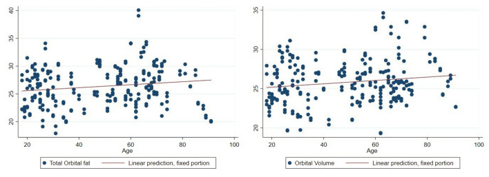
Results: Our findings revealed distinct patterns of age-related changes in the periorbital region. Upper orbital fat remained stable with age, while lower orbital fat exhibited a substantial increase in both anterior and posterior compartments. Notably, orbital cavity volume expanded with bony resorption, while eye globe volume decreased, contributing to an enophthalmic appearance. We observed no vertical displacement of the globe with aging.
Conclusion: This study provides a comprehensive overview of the multifaceted anatomical changes that occur in the periorbital region with aging. The insights gained from this research offer important clinical implications for addressing the signs of periorbital aging, guiding surgical interventions, and ultimately enhancing patient outcomes.
Level of evidence iv: This journal requires that authors assign a level of evidence to each article. For a full description of these Evidence-Based Medicine ratings, please refer to the Table of Contents or the online Instructions to Authors www.springer.com/00266 .
Keywords: Orbital aging; Orbital fat; Periorbital rejuvenation.
© 2025. The Author(s).
Conflict of interest statement
Declarations. Conflict of interest: The authors declare that they have no conflicts of interest to disclose. Human and Animal Rights Statements: This article does not contain any studies with human participants or animals performed by any of the authors. Informed Consent: For this type of study, informed consent is not required.
Figures


Similar articles
-
Periorbital Lipogranuloma Following Facial Autologous Fat Injections: Non-surgical Treatment.Aesthetic Plast Surg. 2015 Dec;39(6):946-52. doi: 10.1007/s00266-015-0554-0. Epub 2015 Aug 26. Aesthetic Plast Surg. 2015. PMID: 26306704
-
Surgical Correction of Tear Trough Deformity (TTD) with Orbicularis Retaining Ligament Release and Volume Augmentation for Periorbital Rejuvenation: Review of the Literature.Aesthetic Plast Surg. 2023 Feb;47(1):199-214. doi: 10.1007/s00266-022-03183-0. Epub 2022 Dec 1. Aesthetic Plast Surg. 2023. PMID: 36456652 Review.
-
Orbital Fat Injection: Technique and 5-Year Follow-Up.Aesthetic Plast Surg. 2019 Feb;43(1):123-132. doi: 10.1007/s00266-018-1205-z. Epub 2018 Sep 21. Aesthetic Plast Surg. 2019. PMID: 30242462
-
Combining High-Density Fat and Condensed Low-Density Fat Injections for Precise Facial Rejuvenation.Aesthetic Plast Surg. 2024 Jun;48(11):2147-2154. doi: 10.1007/s00266-024-03953-y. Epub 2024 Mar 29. Aesthetic Plast Surg. 2024. PMID: 38551708
-
Fat Injection: A Systematic Review of Injection Volumes by Facial Subunit.Aesthetic Plast Surg. 2018 Oct;42(5):1261-1270. doi: 10.1007/s00266-017-0936-6. Epub 2017 Aug 8. Aesthetic Plast Surg. 2018. PMID: 28791455
References
-
- Berger AJ, Kahn D (2012) Growth and development of the orbit. Oral Maxillofac Surg Clin North Am 24(4):545–555 - PubMed
-
- Karunanayake M et al (2017) Analysis of craniofacial remodeling in the aging midface using reconstructed three-dimensional models in paired individuals. Plast Reconstr Surg 140(3):448e–454e - PubMed
-
- Gierloff M et al (2012) Aging changes of the midfacial fat compartments: a computed tomographic study. Plast Reconstr Surg 129(1):263–273 - PubMed
-
- Rohrich RJ, Avashia YJ, Savetsky IL (2021) Prediction of facial aging using the facial fat compartments. Plast Reconstr Surg 147(1s–2):38s–42s - PubMed
MeSH terms
LinkOut - more resources
Full Text Sources
Medical
Research Materials
