

Mapping simulated visual field defects with movie-viewing pupil perimetry
- PMID: 39779511
- PMCID: PMC12238215
- DOI: 10.1007/s00417-024-06733-1
Mapping simulated visual field defects with movie-viewing pupil perimetry
Abstract
Purpose: Assessing the quality of the visual field is important for the diagnosis of ophthalmic and neurological diseases and, consequently, for rehabilitation. Visual field defects (VFDs) are typically assessed using standard automated perimetry (SAP). However, SAP requires participants to understand instructions, maintain fixation and sustained attention, and provide overt responses. These aspects make SAP less suitable for very young or cognitively impaired populations. Here we investigate the feasibility of a new and less demanding form of perimetry. This method assesses visual sensitivity based on pupil responses while performing the perhaps simplest task imaginable: watching movies.
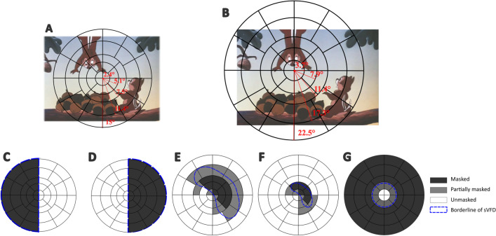
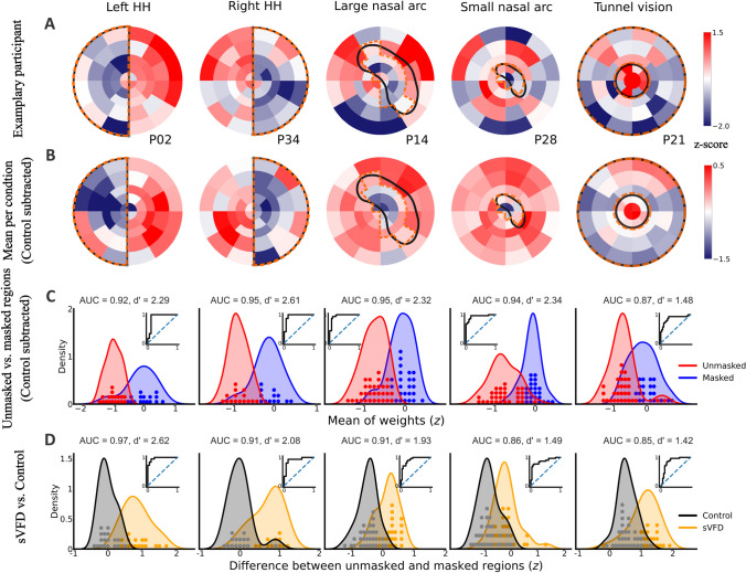
Method: We analyzed an existing dataset, with healthy participants (n = 70) freely watching movies with or without gaze-contingent simulated VFDs, either hemianopia (left- or right-sided) or glaucoma (large nasal arc, small nasal arc, and tunnel vision). Meanwhile, their gaze and pupil size were recorded. Using a recently published toolbox (Open-DPSM), we modeled the relative contribution of visual events to the pupil responses to indicate relative visual sensitivity across the visual field and to dissociate between conditions with and without simulated VFDs.
Result: Conditions with and without simulated VFDs could be dissociated, with an AUC ranging from 0.85 to 0.97, depending on the specific simulated VFD condition. In addition, the dissociation was better when including more movies in the modeling but the model with as few movies as 10 movies was sufficient for a good classification (AUC ranging from 0.84 to 0.96).
Conclusion: Movie-viewing pupil perimetry is promising in providing complementary information for the diagnosis of VFDs, especially for those who are unable to perform conventional perimetry.
Keywords: Glaucoma; Hemianopia; Modeling pupil size; Pupil perimetry; Simulated visual field defects.
© 2025. The Author(s).
Conflict of interest statement
Declarations. Ethical Approval: The data collection was approved by the ethics committee of the Department of Psychology of the University of Groningen (RUG). Informed consent was obtained from all individual participants included in the study. The study followed the tenets of the Declaration of Helsinki. Compliance with ethical Standards: This study was funded by: M. Naber and F.W. Cornelissen were supported by the Algemene Nederlandse Vereniging ter Voorkoming van Blindheid (General Dutch Association for Preventing Blindness) en de Landelijke Stichting voor Blinden en Slechtzienden (National Foundation for the blind and visually impaired) through an UitZicht grant (UZ-2023–18). Y. Cai was supported by a China Scholarship Council (CSC) scholarship. A.F. Ten Brink was funded by the Dutch Research Council (NWO; 406.XS.04.127). Preregistration: The experiment was not preregistered. Conflict of interest: No Conflicts of Interest.
Figures
Similar articles
-
Comparison of Two Modern Survival Prediction Tools, SORG-MLA and METSSS, in Patients With Symptomatic Long-bone Metastases Who Underwent Local Treatment With Surgery Followed by Radiotherapy and With Radiotherapy Alone.Clin Orthop Relat Res. 2024 Dec 1;482(12):2193-2208. doi: 10.1097/CORR.0000000000003185. Epub 2024 Jul 23. Clin Orthop Relat Res. 2024. PMID: 39051924
-
Surgical interventions for bilateral congenital cataract in children aged two years and under.Cochrane Database Syst Rev. 2022 Sep 15;9(9):CD003171. doi: 10.1002/14651858.CD003171.pub3. Cochrane Database Syst Rev. 2022. PMID: 36107778 Free PMC article.
-
Sexual Harassment and Prevention Training.2024 Mar 29. In: StatPearls [Internet]. Treasure Island (FL): StatPearls Publishing; 2025 Jan–. 2024 Mar 29. In: StatPearls [Internet]. Treasure Island (FL): StatPearls Publishing; 2025 Jan–. PMID: 36508513 Free Books & Documents.
-
Cost-effectiveness of using prognostic information to select women with breast cancer for adjuvant systemic therapy.Health Technol Assess. 2006 Sep;10(34):iii-iv, ix-xi, 1-204. doi: 10.3310/hta10340. Health Technol Assess. 2006. PMID: 16959170
-
Comparing a Head-Mounted Smartphone Visual Field Analyzer to Standard Automated Perimetry in Glaucoma: A Prospective Study.J Glaucoma. 2024 Oct 1;33(10):742-747. doi: 10.1097/IJG.0000000000002452. Epub 2024 Jun 17. J Glaucoma. 2024. PMID: 38884623
Cited by
-
Uncovering Distinct Drivers of Covert Attention in Complex Environments With Pupillometry.Psychophysiology. 2025 Mar;62(3):e70036. doi: 10.1111/psyp.70036. Psychophysiology. 2025. PMID: 40104950 Free PMC article.
References
-
- Abeln B, Love R (2018) An Overview of Munchausen Syndrome and Munchausen Syndrome by Proxy. Nurs Clin North Am 53(3):375–384. 10.1016/j.cnur.2018.04.005 - PubMed
-
- Anderson, D. R., & Patella, V. M. (1999). Automated Static Perimetry (2nd edition, Vol. 117). Mosby.
-
- Artes PH, Iwase A, Ohno Y, Kitazawa Y, Chauhan BC (2002) Properties of perimetric threshold estimates from Full Threshold, SITA Standard, and SITA Fast strategies. Invest Ophthalmol Vis Sci 43(8):2654–2659 - PubMed
MeSH terms
Grants and funding
LinkOut - more resources
Full Text Sources
Miscellaneous
