. 2025 Jan 9;124(1):3.
doi: 10.1007/s00436-024-08446-0.

In vitro culture and confocal microscopy study of Maritrema gratiosum Nicoll, 1907 (Digenea): From metacercaria to ovigerous adult

Affiliations

In vitro culture and confocal microscopy study of Maritrema gratiosum Nicoll, 1907 (Digenea): From metacercaria to ovigerous adult

YuChing Chuang et al. Parasitol Res. .

Abstract

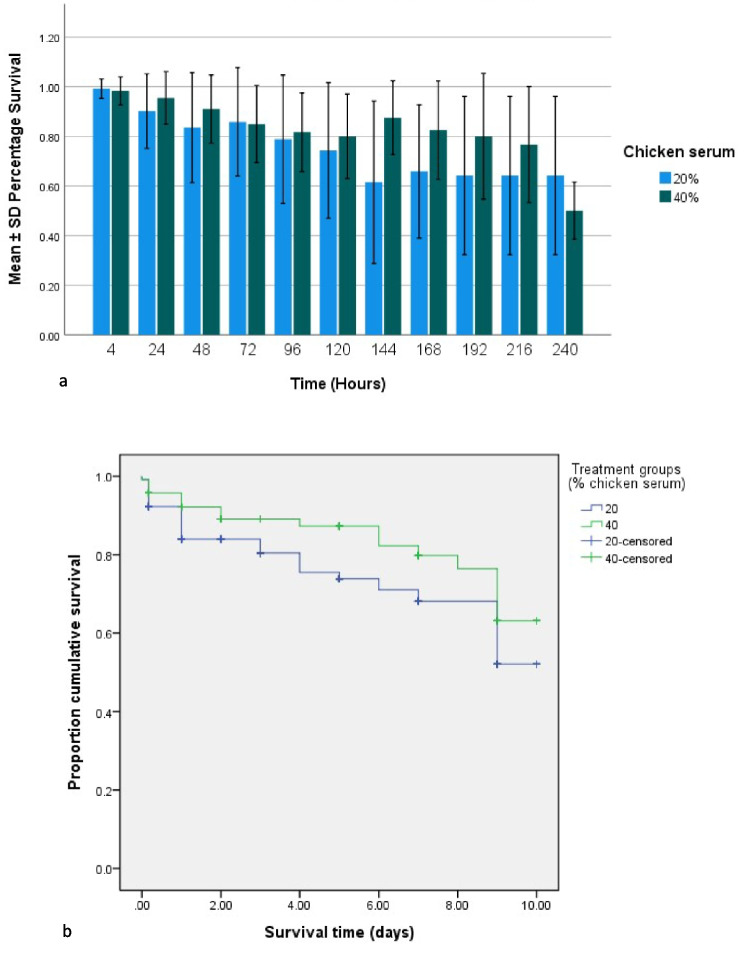
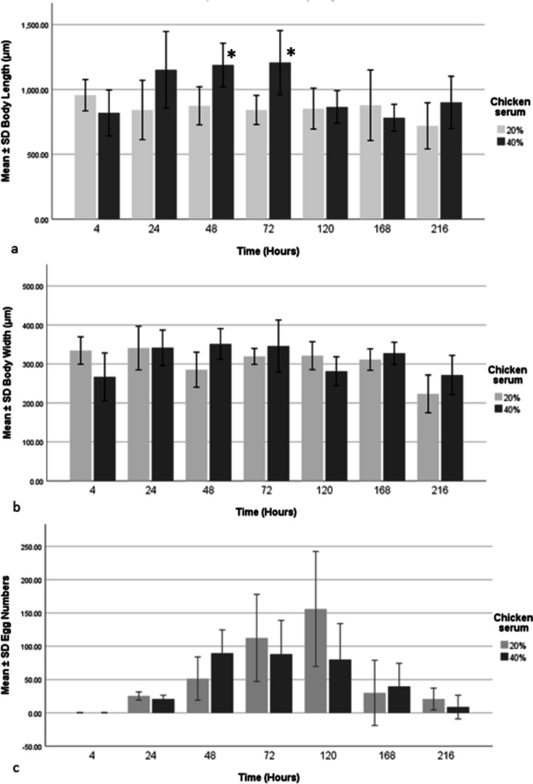
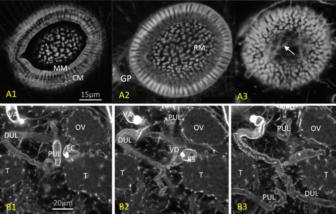
This study set out to characterise the in vitro development, including musculature, of the microphallid parasite of the barnacle Semibalanus balanoides (Linnaeus, 1767), Maritrema gratiosum Nicoll, 1907 collected in Scotland. An in vitro culture model was developed to obtain ovigerous adults of M. gratiosum and their morphology was observed. Different media were tested and NCTC-109 was chosen as the best medium. The effects of different concentrations of serum upon adult longevity, size and egg production was measured. Survival for 10-days was achieved when flukes were cultured in NCTC-109 plus chicken serum and antibiotics. Forty percent chicken serum seemed to provide better results in terms of survival time and producing flukes with the largest body lengths. Both normal and abnormal eggs were observed from adults cultured in vitro. Confocal microscopy was undertaken to provide details of the development of the parasite's ultrastructure, including musculature, during the course of in vitro culture. While the musculature of M. gratiosum was similar to that of other microphallids, some additional novel structures were observed, most notably a ligament connecting pars prostatica and seminal vesicle and a racket-shaped excretory bladder. This study has provided greater insight into the biology M. gratiosum, and also developed a good in vitro model which might be applied to ecological or medical research in the future.

Keywords: Barnacle parasites; Egg production; Excystment rates; Metacercarial development; Musculature; Spermatogenesis.


Conflict of interest statement

Declarations. Ethical approval: No ethical approval was required for the work with barnacles; however, our practices align with ethical principles regarding the collection and treatment of animals and adhere to the animal welfare regulations in the University of Stirling and the UK. Consent to participate: All authors consent to participate in this publication. Consent for publication: All authors consent to publish the manuscript. Competing interests: The authors declare no competing interests. Research data: Available on request by contacting the lead author. Clinical trial: Clinical trial number: not applicable.

Figures

Fig. 1
Fig. 1
Fertilization process. A Note the refractive fertilization chamber between the testis and ovary (30 min in 0.01 M PBS). B Note a spermatozoan (arrow) and an ovum (asterisk) in the fertilization chamber (15 min in 0.01 M PBS). C Note the spermatozoa (arrow) in the seminal vesicle (60 min in 0.01 M PBS). D Spermatozoa in the duct (arrow) between the seminal vesicle and the ductus ejaculatoris (asterisk) (60 min in 0.01 M PBS). FC: fertilization chamber, OV: ovary, T: testis. Figures produced by Olympus cellSens software
Fig. 2
Fig. 2
A Comparison of the mean ± SD percentage survival of excysted distomes of Maritrema gratiosum in NCTC with 20% (+ 100 U ml-1 penicillin + streptomycin 100 μg mL-1) and 40% chicken serum (+ 200 U ml-1 penicillin + streptomycin 200 μg mL-1) over 240 h post-excystment and 40ºC. B Survival curves for the two treatment groups according to the Kaplan–Meier method. Figures produced by IBM SPSS 25
Fig. 3
Fig. 3
Comparison Maritrema gratiosum distome length (A), width (B), and egg numbers (C) at seven time points over 216 h post excystment cultured in NCTC 109 with either 20% (+ 100 U ml-1 penicillin + streptomycin 100 μg mL-1) or 40% chicken serum (+ 200 U ml-1 penicillin + streptomycin 200 μg mL-1) at 40ºC. *Statistically significant differences (p < 0.007) were found in body length. Body length and width were compared using independent sample t-tests with a Bonferroni correction (adjusted significance p < 0.007) to account for multiple comparisons. The figures were produced using IBM SPSS Statistics version 25
Fig. 4
Fig. 4
A Whole worm micrograph, note that the distal uterus (U) is full of eggs (NCTC 109 supplemented with 40% CS and antibiotics for 48 h, ventral view). B Higher magnification showing the male copulatory organs (NCTC 109 supplemented with 20% CS and antibiotics for 24 h). C Higher magnification showing the female copulatory organs (NCTC 109 supplemented with 40% CS and antibiotics for 72 h). D Eggs in the uterus; note the cells at one or both ends of the egg, which are likely vitelline globules, and an ovum in the middle. E Receptaculum seminis and a developing egg inside; note the eggshell and visible Mehlis gland (NCTC 109 supplemented with 20% CS and antibiotics for 24 h). F Higher magnification of the uterus loop; note an ovum and a spermatozoan inside (NCTC 109 supplemented with 20% CS and antibiotics for 72 h). G Lower magnification of the uterus loop; note both normal and abnormal eggs (NCTC 109 supplemented with 20% CS and antibiotics for 168 h). The abnormal eggs looked granular without shells or had deformed shells and had accumulated in the distal part of the uterus. H An adult cultured in NCTC 109 supplemented with 20% CS and antibiotics for 216 h, showing signs of senescence. Whole worm ventral view. CS: cirrus sac. DE: ductus ejaculatoris. E: egg. FC: fertilization chamber. INT: intestine caecum. MEH: Mehlis gland. OS: oral sucker. OV: ovary. OVU: ovum. P: pharynx, PP: pars prostatica. RS: receptaculum seminis, S: spermatozoan, SV: seminal vesicle. T: testis. U: uterus. VA: vagina. VIG: vitelline globules, VS: ventral sucker. Figures produced by Olympus cellSens software
Fig. 5
Fig. 5
Maritrema gratiosum-morphology under LSCM. A DAPI (blue) and phalloidin (green) stain combined white light images (red) (NCTC-109 supplemented with 40% chicken serum and antibiotics for 120 h, dorsal view). Note that the imaged uterus was full of eggs (red), some of which have escaped the uterus due to a fracture in the specimen. B Reproductive organs stained with DAPI (NCTC-109 supplemented with 20% CS and antibiotics for 48 h, ventral view). C Male reproductive system (NCTC-109 supplemented with 40% CS and antibiotics for 120 h, ventral view of anaglyph 3D image). Note the ligament connecting the pars prostatica and seminal vesicle (arrow), showing phalloidin channel only. D. The same specimen as C but showing DAPI channel only. E Tegumental structures showing DAPI channel only (NCTC-109 supplemented with 20% CS and antibiotics for 48 h). Note the “finger-like” structures (arrow) near the surface of the worm. F Excretory pore showing clear radial and circular muscles and two nearby flame cells (NCTC-109 supplemented with 40% CS and antibiotics for 120 h and stained with phalloidin). CS: cirrus sac. DE: ductus ejaculatoris. EP: excretory pore. F: flame cells. GA: genital atrium. INT: intestine caecum. MP: male pore at genital atrium. OE: oesophagus. OS: oral sucker. OV: ovary. PG: prostate glands. P: pharynx. PP: pars prostatica. SV: seminal vesicle. T: testis. U: uterus. VA: vagina. VG: vaginal gland. VI: vitellaria. VS: ventral sucker. Figures produced by the embedded software of confocal microscopy (Leica TCS 2 AOBS LSCM)
Fig. 6
Fig. 6
A Different optical sectioning depths of the ventral sucker of a newly excysted specimen of Maritrema gratiosum that was subsequently stained with × 8 phalloidin. A1. The exterior-most circular and meridial muscles. A2. The radial muscles inside and the genital pore on the side. A3. Extra supportive muscles (arrow) at the base of ventral sucker. B Ootypic junction of a newly excysted M. gratiosum stained with × 8 phalloidin moving from the dorsal side (left) to the ventral side (right). B1. Image showing ovary, fertilisation chamber, proximal uterus loop, distal uterus loop, testis, and vagina. B2. Image showing PUL and DUL, receptaculum seminis, ovary, testes, vagina, and vitelline duct. B3. The duct (asterisk) connecting the fertilisation chamber (can only be seen in B2) and (can only be seen in B1). Figures produced by the embedded software of confocal microscopy (Leica TCS 2 AOBS LSCM). CM: circular muscle. DUL: distal uterus loop. FC: fertilisation chamber. GP: genital pore. MM: meridial muscles. OV: ovary. PUL: proximal uterus loop. RM: radial muscle. RS: receptaculum seminis. T: testis. VA: vagina. VD: vitelline duct
Fig. 7
Fig. 7
Newly excysted specimen of Maritrema gratiosum stained with × 8 phalloidin. Focusing on internal organs of the hind body of a newly excysted M. gratiosum stained with × 8 phalloidin. Dorsal view. The cirrus sac, excretory bladder, intestine caecum, ootypic junction (yellow square), ovary, vagina, ventral sucker, uterus loops and six flame cells (arrows) were clearly visible. Inset figure: Excretory bladder; note the racket-shaped excretory bladder with four side branches. Figures produced by the embedded software of confocal microscopy (Leica TCS 2 AOBS LSCM). CS: cirrus sac. EB: excretory bladder. INT: intestine caecum. OV: ovary. VA: vagina. VS: ventral sucker. UL: uterus loops

Similar articles

References

    1. Abdulla MH, Ruelas DS, Wolff B, Snedecor J, Lim KC, Xu F, Renslo AR, Williams J, McKerrow JH, Caffrey CR (2009) Drug discovery for schistosomiasis: hit and lead compounds identified in a library of known drugs by medium-throughput phenotypic screening. PLoS Negl Trop Dis 3(7):e478. 10.1371/journal.pntd.0000478 - PMC - PubMed
    1. Augot D, Rondelaud D, Dreyfuss G, Cabaret J (1997) Fasciola hepatica: in vitro production of daughter rediae and cercariae from first- and second-generation rediae. Parasitol Res 83:383–385 - PubMed
    1. Basch PF (1981a) Cultivation of Schistosoma mansoni in vitro. I. Establishment of cultures from cercariae and development until pairing. J Parasitol 67(2):179–185 - PubMed
    1. Basch PF (1981b) Cultivation of Schistosoma mansoni in vitro. II. Production of infertile eggs by worm pairs cultured from cercariae. J Parasitol 67(2):186–190 - PubMed
    1. Benjamin LR, James BL (1987) The development of the metacercariae of Maritrema linguilla Jäg., 1908 (Digenea: Microphallidae) in the intermediate host, Ligia oceanica (L.). Parasitology 94:221–231

LinkOut - more resources