. 2025 Jan 8;16(1):513.
doi: 10.1038/s41467-024-55544-9.

CCR5-ligand decorated rilpivirine lipid-based nanoparticles for sustained antiretroviral responses

Affiliations

CCR5-ligand decorated rilpivirine lipid-based nanoparticles for sustained antiretroviral responses

Milankumar Patel et al. Nat Commun. .

Abstract

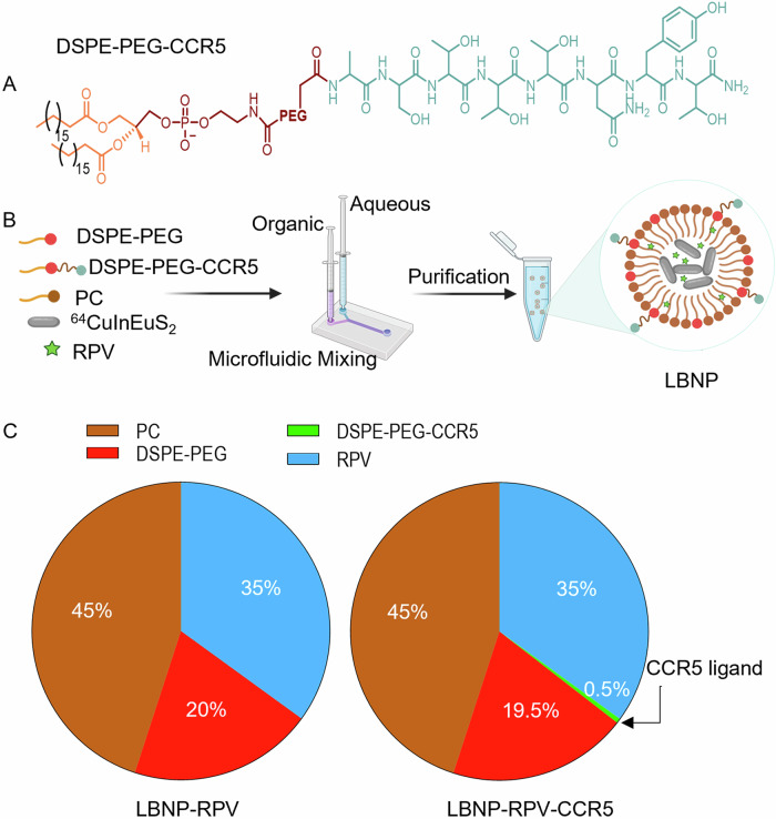
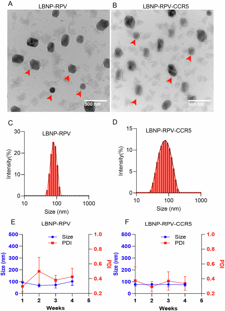
Antiretroviral therapy (ART) improves the quality of life for those living with the human immunodeficiency virus type one (HIV-1). However, poor compliance reduces ART effectiveness and leads to immune compromise, viral mutations, and disease co-morbidities. Here we develop a drug formulation in which a lipid-based nanoparticle (LBNP) carrying rilpivirine (RPV) is decorated with the C-C chemokine receptor type 5 (CCR5) targeting peptide. This facilitates extended drug persistence within myeloid cells. Particle delivery to viral reservoirs is tracked by positron emission tomography. The CCR5-mediated LBNP cell uptake and retention reduce HIV-1 replication in human monocyte-derived macrophages and infected humanized mice (hu mice). Focused ultrasound with microbubbles mediated blood brain barrier (BBB) disruption allows the CCR5-targeted LBNP to penetrate the BBB and reach brain myeloid cells. These findings offer a role for CCR5-targeted therapeutics in antiretroviral delivery to optimize HIV suppression.


Conflict of interest statement

Competing interests: The authors declare that Dr. Howard Gendelman co-founder of Exavir Therapeutics, Inc. The biotechnology company is developing ultra-long-acting drugs. The drugs in development are not linked to those created in the current report. All other authors declare no competing interests.

Figures

Fig. 1
Fig. 1. Morphological characterization of the multimodal CuInEuS2 nanoprobe.
A, B Transmission electron microscope images of the multimodal rod-shaped nanoprobes. CuInEuS2 and its crystal lattice (red lines) are illustrated. C A scanning transmission electron microscopy (STEM) map shows elemental localization within the nanoprobe from a corresponding high-angle annular dark-field electron microscopy image. The map shows the presence of copper (red), indium (blue), europium (cyan), and sulfur (green) in the nanoprobe. The reproducibility of CuInEuS2 synthesis was verified at least 10 independent reactions. D XRD pattern of CuInEuS2 nanoprobe demonstrates a crystal structure.
Fig. 2
Fig. 2. Synthesis, formulation, and composition of LBNPs.
A The chemical structure of the DSPE-PEG conjugated CCR5-receptor targeting peptide is shown. B Schematic illustration of the formulation of LBNPs by microfluidic mixing, created in BioRender. C The molar percentages of various lipids used in LBNP formulation are presented in the pie chart.
Fig. 3
Fig. 3. Physicochemical LBNP characterization.
A, B The TEM image shows a spherical LBNP-RPV and LBNP-RPV-CCR5 morphology. The red arrowheads were used to indicate the representative particles. C, D The DLS size profile demonstrates the unimodular distribution of the LBNPs. E, F The changes in size and polydispersity of the LBNPs were recorded for one month at 4 °C. LBNPs were stable without changes in size and polydispersity. Results are expressed as the mean ± SD for independent replicates (n = 3).
Fig. 4
Fig. 4. CCR5–receptor-induced LBNPs uptake, depot formation and HIV-1 viral suppression in MDMs, Ex-vivo.
A Dose-associated macrophage viability measurements following LBNP exposures by the CTB assay after 24 h incubation. Both the LBNP-RPV and LBNP-RPV-CCR5 showed cell viability above 90% up to 100 μM RPV concentration. B LBNP-RPV and LBNP-RPV-CCR5 macrophage uptake was analyzed by measuring RPV concentration for 24 h at 20 μM RPV. LBNP-RPV-CCR5 showed a 3-fold increase in drug uptake than LBNP-RPV. C The CCR5 inhibitor (maraviroc, 1 nM) attenuated the uptake of LBNP-RPV-CCR5. D Confocal microscopy was performed with Cy5.5 dye-labeled LBNP-RPV-CCR5 treated macrophages in the presence (lower panel) and absence (upper panel) of maraviroc. The nuclei and cell membranes were stained with DAPI (blue, stained DNA) and phalloidin (green, stained F-actin), respectively. E RPV retention was evaluated by measuring RPV in the cell supernatant fluids. F TEM image of LBNP engulfed macrophage. Macrophages show multiple depots in LBNP-RPV-CCR5 than in LBNP-RPV as highlighted by red arrowhead. G The viral suppression in LBNP-RPV and LBNP-RPV-CCR5 treated macrophages was evaluated by measuring virus levels in the cell supernatant fluids. The data were collected over 25 days after a 100 μM administered dose. The reproducibility of cellular uptake and depot formation were verified by at least 3 independent experiments. All results are expressed as the mean ± SD for n = 3 biological replicates. Statistical analysis was performed by an unpaired t-test. ****, P < 0.0001; and ns = not significant.
Fig. 5
Fig. 5. LBNP and RPV tissue biodistribution in mice by PET imaging and mass spectrometry.
A Schematic presentation of LBNP biodistribution by PET. B Humanized mice were injected with LBNP-RPV or LBNP-RPV-CCR5, and particle biodistribution was monitored by PET at 6, 24, and 48 h. Both coronal (left panel) and sagittal (right panel) views of mice are illustrated. C, D Quantitative measurements of radiolabeled LBNPs in tissue were recorded at 48 h post-administration by using a gamma counter. LBNP-RPV-CCR5 was concentrated in the spleen. E Spleen/liver radioactivity ratios for LBNP-RPV-CCR5 and LBNP-RPV are shown. F A schematic presentation of LBNP injection and RPV quantitation in humanized mice is shown. G Plasma RPV levels in mice treated with LBNP-RPV and LBNP-RPV-CCR5 are shown at 6 and 24 h post-injection. H The spleen/liver RPV ratio at 24 h post-injection of LBNPs. The results presented in Figures A to E were obtained with nanoprobe-containing LBNPs, whereas those in Figures F to H were acquired using LBNPs without the nanoprobe. Figures A and F were created in BioRender. Results are presented as the mean ± SD for n =  3 biological replicates. Statistical significance was calculated by using an unpaired t-test, **P < 0.01, and ****P < 0.0001.
Fig. 6
Fig. 6. Brain delivery of LBNP-RPV-CCR5 nanoparticles.
A Schematic presentation of FUS-treated humanized microglial (MG) mice that received LBNP treatment, created in BioRender. B IVIS images show bright yellow Cy5.5 signals in the brains of FUS-treated mice (middle) that received LBNP-RPV-CCR5, compared to those without FUS treatment (left). With FUS treatment, the mice receiving LBNP-RPV-CCR5 (middle) show higher brain accumulation than those receiving LBNP-RPV (right). C Both mice showed good BBB disruption, as shown by the gadolinium enhancements (bright signals, blue arrows) on the coronal sections of the T1-weighted images (T1WI) on MRI. The susceptibility-weighted imaging (SWI) revealed the absence of microhemorrhages. D The brain RPV level in LBNP-RPV-CCR5 treated mice with and without FUS. E The fluorescence microscopy image of brain tissue sections, stained with IBA-1 (red, microglia) and HuNu (green, human nuclei). Approximately 50% of microglia (IBA-1, red stain) in the hu mice were positive for the human marker (HuNu, green stain, white arrows), while mouse microglia are not immunoreactive to HuNu (blue arrows). Both the LBNP-RPV and LBNP-RPV-CCR5 treated mice show Cy5.5 signals in the brain tissue; however, only the microglia with the HuNu stains showed engulfment of the LBNPs as the Cy5.5 signals appeared in the cytoplasm of the human microglia (white arrows). F Quantification of Cy5.5 signal intensity from fluorescence microscopy images of brain tissue, analyzed using ImageJ. Furthermore, the Cy5.5 signals appear to be more intense in the animals that received the LBNP-RPV-CCR5 than LBNP-RPV. Scale bar= 25 µm. All results are expressed as the mean ± SD for n = 3 biological replicates. Statistical significance was calculated using an unpaired t-test, *P < 0.05 and ****P < 0.0001.
Fig. 7
Fig. 7. Viral suppression and toxicity profiles of RPV-encapsulated LBNP.
A Schematic representations of experimental timelines are shown for LBNP treatments, Created in BioRender. B Viral suppression efficiency of LBNP-RPV and LBNP-RPV-CCR5 on day 14 are illustrated. LBNP-RPV-CCR5 showed complete viral suppression in 2/3 of the treated mice up to day 14 (number animals n = 3 per group). C The change in mice body weight in the untreated, LBNP-RPV, and LBNP-RPV-CCR5 treated group. D The histological images of hematoxylin and eosin-stained heart, lung, kidney, spleen, liver, and brain tissue sections from the treatment groups. The images were captured at 20X magnification (Scale bar 200 µm). All results are expressed as the mean ± SD for n = 3 biological replicates. Statistical significance was calculated using an unpaired t-test, *P < 0.05.

Update of

References

    1. Khan, N. H. et al. Assessment of knowledge and attitudes related to hiv/aids among the population with increasing incidence rate. Cureus16, e53451 (2024). - PMC - PubMed
    1. Bekker, L.-G. et al. HIV infection. Nat. Rev. Dis. Prim.9, 42 (2023). - PubMed
    1. Holmes, E. C. On the origin and evolution of the human immunodeficiency virus (HIV). Biol. Rev.76, 239–254 (2001). - PubMed
    1. Chun, T.-W., Moir, S. & Fauci, A. S. HIV reservoirs as obstacles and opportunities for an HIV cure. Nat. Immunol.16, 584–589 (2015). - PubMed
    1. Senapathi, J., Bommakanti, A., Mallepalli, S., Mukhopadhyay, S. & Kondapi, A. K. Sulfonate modified Lactoferrin nanoparticles as drug carriers with dual activity against HIV-1. Colloids Surf. B Biointerfaces191, 110979 (2020). - PubMed

MeSH terms

LinkOut - more resources