. 2025 Jan 8;15(1):1283.
doi: 10.1038/s41598-024-82121-3.

SIX1 aggravates the progression of spinal cord injury in mice by promoting M1 polarization of microglia

Affiliations

SIX1 aggravates the progression of spinal cord injury in mice by promoting M1 polarization of microglia

Zhonghua Xu et al. Sci Rep. .

Abstract

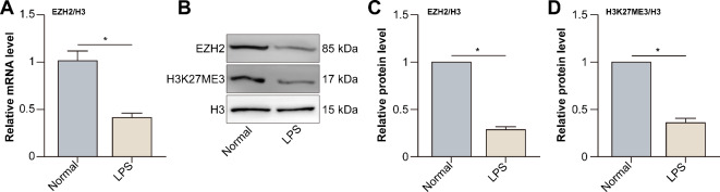
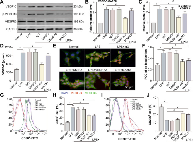
Inflammation aggravates secondary damage following spinal cord injury (SCI). M1 microglia induce inflammation and exert neurotoxic effects, whereas M2 microglia exert anti-inflammatory and neuroprotective effects. The sine oculis homeobox (SIX) gene family consists of six members, including sine oculis homeobox homolog 1 (SIX1)-SIX6. SIX1 is expressed in microglia and promotes inflammation. This study aimed to evaluate the role and underlying mechanisms of SIX1 in microglia polarization in vitro (LPS-treated mouse microglia; BV2 cells) and in vivo (a mouse model of SCI). SIX1 expression was increased in the microglia of mice with SCI. SIX1 was positively correlated with the M1 microglia marker inducible nitric oxide synthase (iNOS) and negatively correlated with the M2 microglia marker arginase 1 (Arg1) in mice with SCI. Knockdown of SIX1 promoted functional recovery by enhancing M2 microglia polarization in mice with SCI. The transcription, expression, and activity of enhancer of zeste homolog 2 (EZH2) were decreased in LPS-stimulated BV2 cells. Downregulation of EZH2 promoted SIX1 expression in LPS-treated BV2 cells by inhibiting the methylation of the SIX1 promoter. SIX1 enhanced the transcription of vascular endothelial growth factor-C (VEGF-C) in LPS-stimulated BV2 cells with downregulated EZH2. VEGF-C promoted M1 polarization and inhibited M2 polarization in BV2 cells by binding to vascular endothelial growth factor receptor 3 (VEGFR3). Overall, the results suggest that SIX1 promotes M1 polarization of microglia following SCI by upregulating the VEGF-C/VEGFR3 axis, whereas the blockade of SIX1 can improve the recovery of locomotor function following SCI, demonstrating a novel strategy for the treatment of SCI.

Keywords: Microglia; Polarization; Sine oculis homeobox homolog 1 (SIX1); Spinal cord injury (SCI); Vascular endothelial growth factor-C (VEGF-C).


Conflict of interest statement

Declarations. Competing interests: The authors declare no competing interests. Ethical approval: We confirm that all experimental protocols conducted in this study were approved by the Medical Ethics Committee of the Third Affiliated Hospital of Soochow University. We confirm that all methods used in this study were carried out in accordance with relevant guidelines and regulations. The study was carried out in compliance with the ARRIVE guidelines.

Figures

Fig. 1
Fig. 1
SIX1 expression in microglia increases following SCI in mice. (A) Mice were divided into the normal, sham, SCI-3-d, and SCI-7-d groups. HE staining of longitudinal sections of mouse spinal cords was shown. The areas around dotted lines represented injury spinal cords. Mice were divided into the normal, sham, SCI-1-d, SCI-3-d, SCI-5-d, SCI-7-d, SCI-14-d, SCI-21-d, SCI-28-d, and SCI-35-d groups. (B) Western blotting was performed to examine the expression of SIX1, SIX2, SIX3, and SIX6 in the spinal cord of mice. (CH) The expression of SIX1, SIX2, SIX3, and SIX6 was evaluated relative to that of β-actin. (I) NeuN and SIX1 were labelled on the spinal cord cross-section of mice in the SCI-7-d group. (J) GFAP and SIX1 were labelled on the spinal cord cross-section of mice in the SCI-7-d group. (K) Iba1 and SIX1 were labelled on the spinal cord cross-section of mice in the SCI-7-d group. (L) Co-localization of SIX1 and NeuN, GFAP, or Iba1. Double staining for SIX1 and NeuN (M), GFAP (N), or Iba1 (O) in the spinal cord cross-sections of mice in the SCI-7-d group. P Co-localization of SIX6 and NeuN, GFAP, or Iba1 (*P < 0.05; **P < 0.01; ***P < 0.001 compared with the normal group; n = 5 per condition). The arrows in (I) indicated the co-localization of Iba1 and SIX1.
Fig. 2
Fig. 2
SIX1 was positively correlated with the M1 microglia marker iNOS and negatively correlated with the M2 microglia marker Arg-1 following SCI. Mice were divided into the normal, sham, SCI-1-d, SCI-3-d, SCI-5-d, SCI-7-d, SCI-14-d, SCI-21-d, SCI-28-d, and SCI-35-d groups. (A) Western blotting was performed to examine the expression of iNOS and Arg-1. (B,C) The expression of iNOS and Arg-1 was analyzed relative to that of β-actin. (D) Pearson correlation analysis was performed to examine the correlation between the expression of SIX1 and iNOS. (E) Pearson correlation analysis was performed to examine the correlation between the expression of SIX1 and Arg-1 (*P < 0.05; **P < 0.01; ***P < 0.001 compared with the normal group; n = 5 per condition).
Fig. 3
Fig. 3
Knockdown of SIX1 promotes locomotor function recovery by enhancing M2 microglia polarization in mice with SCI. Mice were divided into the normal, sham, SCI-35-d, SCI-35-d + lenti-scrambled shRNA, SCI-35-d + lenti-SIX1 shRNA, SCI-35-d + lenti-SIX1 shRNA + LPS + IFN-γ, and SCI-35-d + IL-4 + IL-13 groups. (A) Western blotting was performed to examine SIX1 expression. (B) SIX1 expression relative to GAPDH expression. (C) Gating strategy for detecting M1 microglia (CD11+CD45intCD86+) and M2 microglia (CD11+CD45intCD206+). In the histograms, the red regions represent unstained cells (negative control), and the purple regions on the right of the vertical black lines represent APC-positive (M1) or FITC-positive (M2) cells. The proportions of M1 (D) and M2 (E) microglia in different groups of mice. (F) BMS scores of mice. (G) The time point was set to 35 d post-injury. For footprint analysis in each group, the forelimbs and hindlimbs of rats were immersed in red and blue dyes, respectively. (H) Stride length (*P < 0.05 compared with the normal group; #P < 0.05 compared with the SCI-35-d group; %P < 0.05 compared with the SCI-35-d + SIX1 shRNA group; n = 5 per condition).
Fig. 4
Fig. 4
The transcription, expression, and activity of EZH2 are decreased in LPS-stimulated BV2 cells. BV2 cells were divided into the normal and lipopolysaccharide (LPS) (1 µg/mL for 24 h) groups. (A) qRT-PCR was performed to assess the mRNA expression of EZH2. (B) Western blotting was performed to examine EZH2 and H3K27me3 levels in microglia. Histone 3 (H3) was used as the loading control. (C) EZH2 expression relative to H3 expression. (D) H3K27me3 expression relative to H3 expression (*P < 0.05 compared with the normal group; n = 4 per condition).
Fig. 5
Fig. 5
Downregulated EZH2 promotes SIX1 expression in LPS-treated BV2 cells by inhibiting methylation of the SIX1 promoter. BV2 cells were divided into the normal, LPS, LPS + dCas9 plasmid, and LPS + full-length EZH2-dCas9 groups. (A) qRT-PCR was performed to assess the mRNA expression of SIX1. (B) Western blotting was performed to assess the levels of EZH2, H3K27me3, and SIX1. (CE) Levels of EZH2, SIX1, and H3K27me3 levels relative to those of H3. (F,G) CO-IP was performed to detect the binding of EZH2 to H3K27M. (H) CHIP-qPCR was performed to detect H3K27me3 in the SIX1 promoter. qPCR was performed with primers specific for the SIX1 promoter (*P < 0.05 compared with the normal group; #P < 0.05 compared with the LPS group; n = 4 per condition).
Fig. 6
Fig. 6
SIX1 enhances the transcription of VEGF-C in LPS-stimulated BV2 cells by downregulating EZH2. BV2 cells were divided into the normal, LPS, LPS + lenti-scrambled shRNA, LPS + lenti-SIX1 shRNA, and LPS + lenti-SIX1 shRNA + GSK126 groups. (A) qRT-PCR was performed to assess the mRNA expression of VEGF-C. (B) Western blotting was performed to assess the protein expression of VEGF-C. (C) VEGF-C protein levels relative to GAPDH protein levels. (D) SIX1-binding sites in the VEGF-C promoter. (E) Luciferase reporter assay was performed to detect the activity of the VEGF-C promoter. (F) CHIP was performed to detect the binding of SIX1 to the VEGF-C promoter. qPCR was performed with primers specific for the VEGF-C promoter (*P < 0.05 compared with the normal group; #P < 0.05 compared with the LPS group; %P < 0.05 compared with the LPS + SIX1 shRNA group; n = 4 per condition).
Fig. 7
Fig. 7
VEGF-C promotes M1 polarization and inhibits M2 polarization in BV2 cells by binding to VEGFR3. BV2 cells were divided into the normal, LPS, LPS + rabbit IgG, LPS + 0.01% DMSO, LPS + VEGF-C neutralizing antibody, and LPS + MAZ51 groups. (A) Western blotting was performed to assess the protein levels of VEGF-C, p-VEGFR3, and VEGFR3. (B) VEGF-C protein levels relative to GAPDH protein levels. (C) p-VEGFR3 expression relative to VEGFR3 expression. (D) ELISA was performed to assess the protein levels of VEGF-C in microglia culture supernatant. (E) Double staining for VEGF-C and VEFGR3 was performed to detect their co-localization. (F) Co-localization of VEGF-C and VEGFR3. (G) Flow cytometry was performed to assess the polarization of M1 microglia using the CD86 antibody. (H) Proportion of CD86-positive microglia. (I) Flow cytometry was performed to assess the polarization of M2 microglia using the CD206 antibody. (J) Proportion of CD206-positive microglia (*P < 0.05 compared with the normal group; #P < 0.05 compared with the LPS group; n = 4 per condition).
Fig. 8
Fig. 8
Schematic representation of the mechanism of action of SIX1 in SCI.

Similar articles

References

    1. Ding, W. et al. Spinal cord Injury: The Global incidence, prevalence, and disability from the global burden of Disease Study 2019. Spine (Phila Pa. 1976). 47, 1532–1540 (2022). - PMC - PubMed
    1. Mishra, M. K. et al. Harnessing the benefits of Neuroinflammation: Generation of Macrophages/Microglia with Prominent Remyelinating Properties. J. Neurosci.41, 3366–3385 (2021). - PMC - PubMed
    1. Coppenrath, K. et al. Generation of a new six1-null line in Xenopus tropicalis for study of development and congenital disease. Genesis59, e23453 (2021). - PMC - PubMed
    1. Hsu, J. Y. et al. SIX1 reprograms myogenic transcription factors to maintain the rhabdomyosarcoma undifferentiated state. Cell. Rep.38, 110323 (2022). - PMC - PubMed
    1. Gao, X. et al. Detrimental role of SIX1 in hepatic lipogenesis and fibrosis of non-alcoholic fatty liver disease. Liver Int.43, 1473–1485 (2023). - PubMed

Publication types

MeSH terms