. 2025 Jan 8;15(1):1357.
doi: 10.1038/s41598-024-81508-6.

Novel primers drive accurate SYBR Green PCR detection of Listeria monocytogenes and Listeria innocua in cultures and mushrooms

Affiliations

Novel primers drive accurate SYBR Green PCR detection of Listeria monocytogenes and Listeria innocua in cultures and mushrooms

Bo-Eun Kim et al. Sci Rep. .

Abstract

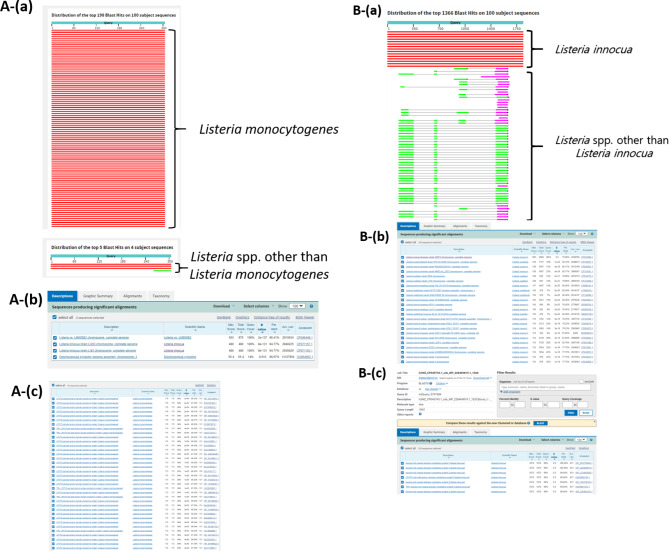
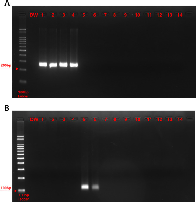
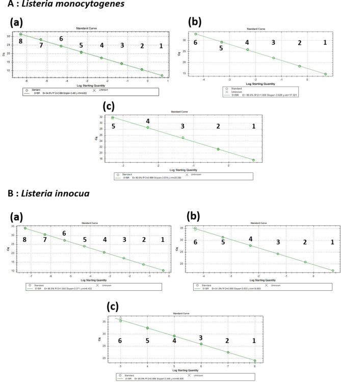
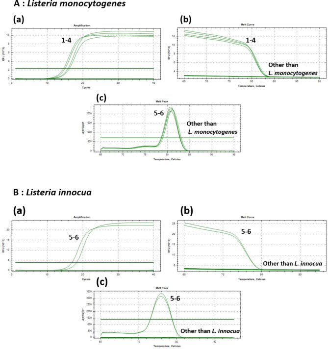
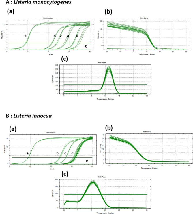
The close genetic resemblance between Listeria monocytogenes and Listeria innocua, combined with their presence in similar environments, poses challenges for species-specific detection in food products. Ensuring food safety through microbiological standards necessitates reliable detection of pathogens like L. monocytogenes and L. innocua throughout the food chain using appropriate analytical techniques. This study aims to develop, identify, and validate a SYBR Green qPCR-based genetic marker designed to detect L. monocytogenes and L. innocua. By performing a comparative analysis of the complete genome sequences of L. monocytogenes (ATCC 12392) and L. innocua (CFSAN044836), a unique gene region encoding a hypothetical protein with an LPXTG cell wall anchor domain (GCF_003031895.1) in L. monocytogenes and leucine-rich repeats (GCF_009648575.1) in L. innocua was identified. Primers targeting these specific region were designed and validated for their effectiveness in detecting L. monocytogenes/L. innocua using both conventional PCR and qPCR techniques. These primers exhibited high sensitivity and specificity in amplifying L. monocytogenes and L. innocua among different Listeria species. The sensitivity and specificity of the primers were further confirmed through standard curve analysis using three different templates: cloned DNA (as a positive control), genomic DNA, and bacterial cell suspension. Additionally, the primers were rigorously tested and validated for their accuracy in directly detecting the targeted strains in live enoki mushroom samples. This direct qPCR method offers significant advantages for the rapid and precise detection of L. monocytogenes and L. innocua, potentially enhancing the efficiency of diagnostic and monitoring processes within food and vegetable distribution systems.

Keywords: L. Innocua; L. monocytogenes; Detection; Primer design; Quantification; qPCR.


Conflict of interest statement

Declarations. Competing interests: The authors declare no competing interests.

Figures

Fig. 1
Fig. 1
(A) L. monocytogenes. (a) Graphic summary of the top 198 BLASTn hits in the target sequence and BLASTn hits excluding L. monocytogenes. (b) Description of BLASTn hits excluding L. monocytogenes. (c) BLASTn of gene amplification region using L. monocytogenes-specific primers. (B) L. innocua. (a) Graphic summary of the top 136 BLASTn hits in the target sequence and BLASTn hits excluding L. innocua. (b) Description of BLASTn hits excluding L. innocua. (c) BLASTn of gene amplification region using L. innocua-specific primers.
Fig. 2
Fig. 2
PCR amplification of L. monocytogenes and L. innocua with the LX250 F/R and INO117 F/R primer sets. (A) PCR amplification of a virtual protein gene (GCF_ 003031895.1) in L. monocytogenes using LX250 F/R primer set. (B) PCR amplification of a virtual protein gene (GCF_009648575.1) in L. innocua using B. INO117 F/R primer set. Lane M: a size marker (100 bp DNA ladder), Lane 1–4: L. monocytogenes strains, Lane 5–6: L. innocua strains, Lane 7–14: other Listeria strains, DW: negative control (distilled water). Details about the strains are shown in Table 1.
Fig. 3
Fig. 3
Standard curve derived from qPCR analysis with LX250 F/R and INO117 F/R primers (A) L. monocytogenes. (a) Cloned DNA: samples 1 to 8 correspond to concentrations ranging from 500 ag/µL to 5 ng/µL. (b) Genomic DNA: samples 1 to 6 correspond to concentrations ranging from 500 ag/µL to 5 ng/µL. (c) Bacterial cell suspensions: Samples 1 to 6 correspond to concentrations ranging from 1.0 × 102 to 1.0 × 109 CFU/mL. (B) L. innocua. (a) Cloned DNA: Samples 1 to 8 correspond to concentrations ranging from 50 ag/µL to 5 ng/µL. (b) Genomic DNA: Samples 1 to 6 correspond to concentrations ranging from 50 ag/µL to 5 ng/µL. (c) Bacterial cell suspensions: Samples 1 to 6 correspond to concentrations ranging from 2.4 × 100 to 2.4 × 10⁸ CFU/mL.
Fig. 4
Fig. 4
Specificity, melting peak, and melting curve of the LX250 F/R and INO117 F/R primer set by SYBR Green qPCR. 5 ng of bacterial genomic DNA was used as the template for qPCR. (A) Detection of L. monocytogenes using the LX250 F/R primer set. (B) Detection of L. innocua using the INO117 F/R primer set. (a) Amplification plot of qPCR. (b) Melt curve of qPCR. (c) Melt peak showing the amplified product’s melting temperature (Tm) of approximately 81 °C. Lanes 1–4: L. monocytogenes strains. Lanes 5–14: Other Listeria strains (described in Table 1). Lane 15: Negative control (distilled water).
Fig. 5
Fig. 5
Species-Specific Detection of Listeria monocytogenes and Listeria innocua in live enoki mushroom samples via Direct SYBR green qPCR analysis. (A) L. monocytogenes. (a) Amplification curves. (b) Melting curves. (c) Melting peaks, confirming that the target gene was amplified at a specific temperature. Peak No 1 Positive control (cloned DNA sample, 5 ng). Peak Nos 2–6. Samples inoculated with Listeria monocytogenes at concentrations ranging from 1.0 × 103 to 1.0 × 109 CFU/mL. (7) Sterile water (negative control). (B) L. innocua. (a) Amplification curves. (b) Melting curves. (c) Melting peaks, confirming that the target gene was amplified at a specific temperature. Peak No 1 Positive control (cloned DNA sample, 5 ng). Peak Nos 2–6. Samples inoculated with Listeria monocytogenes at concentrations ranging from 1.2 × 103 to 1.2 × 109 CFU/mL and non-inoculated enoki mushroom. (5) Sterile water (negative control). Each condition was tested in three biological replicates.

Similar articles

References

    1. Lourenco, A., Linke, K., Wagner, M. & Stessl, B. The saprophytic lifestyle of Listeria monocytogenes and entry into the food-processing environment. Front. Microbiol.13, 789801 (2022). - PMC - PubMed
    1. Rodríguez-Lázaro, D. et al. Quantitative detection of Listeria monocytogenes and Listeria innocua by real-time PCR: Assessment of hly, iap, and lin02483 targets and AmpliFluor technology. Appl. Environ. Microbiol.70(3), 1366–1377 (2004). - PMC - PubMed
    1. Vázquez-Boland, J. A. et al. Listeria pathogenesis and molecular virulence determinants. Clin. Microbiol. Rev.14 (3), 584–640 (2001). - PMC - PubMed
    1. Rodriguez, C., Taminiau, B., García-Fuentes, E., Daube, G. & Korsak, N. Listeria monocytogenes dissemination in farming and primary production: Sources, shedding and control measures. Food Control120, 107540 (2021).
    1. Oevermann, A., Zurbriggen, A. & Vandevelde, M. Rhombencephalitis caused by Listeria monocytogenes in humans and ruminants: A zoonosis on the rise? Interdiscip. Perspect. Infect. Dis.2010(1), 632513 (2010). - PMC - PubMed

Publication types

LinkOut - more resources