. 2025 Jan;637(8047):998-1004.
doi: 10.1038/s41586-024-08355-3. Epub 2025 Jan 8.

Conformational protection of molybdenum nitrogenase by Shethna protein II

Affiliations

Conformational protection of molybdenum nitrogenase by Shethna protein II

Philipp Franke et al. Nature. 2025 Jan.

Abstract

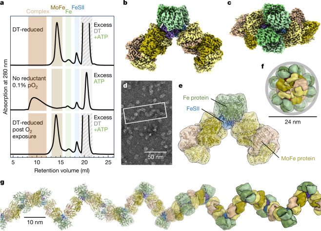
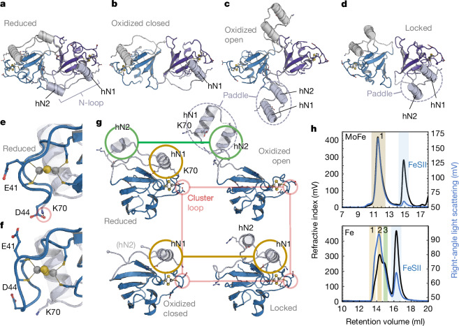
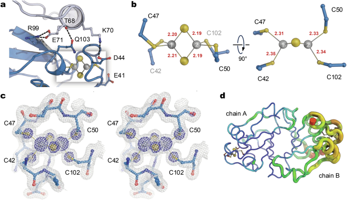
The oxygen-sensitive molybdenum-dependent nitrogenase of Azotobacter vinelandii is protected from oxidative damage by a reversible 'switch-off' mechanism1. It forms a complex with a small ferredoxin, FeSII (ref. 2) or the 'Shethna protein II'3, which acts as an O2 sensor and associates with the two component proteins of nitrogenase when its [2Fe:2S] cluster becomes oxidized4,5. Here we report the three-dimensional structure of the protective ternary complex of the catalytic subunit of Mo-nitrogenase, its cognate reductase and the FeSII protein, determined by single-particle cryo-electron microscopy. The dimeric FeSII protein associates with two copies of each component to assemble a 620 kDa core complex that then polymerizes into large, filamentous structures. This complex is catalytically inactive, but the enzyme components are quickly released and reactivated upon oxygen depletion. The first step in complex formation is the association of FeSII with the more O2-sensitive Fe protein component of nitrogenase during sudden oxidative stress. The action of this small ferredoxin represents a straightforward means of protection from O2 that may be crucial for the maintenance of recombinant nitrogenase in food crops.


Conflict of interest statement

Competing interests: The authors declare no competing interests.

Figures

Fig. 1
Fig. 1. Structure of the O2-protected FeSII–nitrogenase complex.
a, Complex formation monitored by SEC detected by absorption at 280 nm. In a dithionite (DT)-reduced sample (top), the nitrogenase component proteins and FeSII elute as separate peaks without detectable complex formation. Upon removal of reductant and addition of 0.1 vol% O2, complex formation is observed. Reduction and O2 removal lead to complex dissociation (bottom). b, Cryo-EM map of a particle consisting of two copies each of the MoFe protein NifDK and the Fe protein NifH held together by a dimer of the ferredoxin FeSII. c, Top view of the complex shown in a. FeSII is fully buried, and the entire particle follows the C2 symmetry of the ferredoxin. d, Negative-stain TEM image of a representative complex preparation. The assembly forms filaments of variable length and the refined particle structures are sub-averages of such filaments. e, Architecture of the FeSII-protected complex with labelled components. Each protomer of the small FeSII dimer directly contacts one NifH dimer and one MoFe protein. f, Filament formation occurs following the C2 symmetry of MoFe protein, leading to an extended structure with a diameter of 24 nm. g, The filament forms a right-handed helix with a pitch of approximately 30 nm, corresponding to about 1 MDa per helical turn. Scale bars, 50 nm (d), 24 nm (f), 10 nm (g).
Fig. 2
Fig. 2. Conformational states of FeSII.
a, Crystal structure of the reduced FeSII dimer viewed along the C2 axis, with the N-loops shown in grey and light blue. b, Closed dimer in the oxidized structure (PDB 5FFI). c, In the oxidized open state (PDB 5FFI), helices hN1 and hN2 of the N-loop extend outward, forming a paddle-like structure. d, As FeSII forms a complex with the nitrogenase component proteins, the N-loops in this locked state are retracted but the paddle arrangement is retained. hN1 interacts predominantly with NifH, while hN2 interacts with MoFe protein. e, In the reduced state, the cluster-binding loop P39–C50 faces inward, with K70 forming a hydrogen bond to D44. f, In all other structures, the cluster-binding loop faces outward, breaking the D44–K70 interaction and making E41 available to interact with Fe protein (Fig. 3f). g, Monomer structures of the four observed states. Helix hN1 remains close to the [2Fe:2S] cluster in the reduced and locked states, and also in the closed oxidized states, although the H-bond to K70 is broken (brown). Helix hN2 is disordered in the closed oxidized state, but moves only slightly between the reduced and open oxidized state (green). The cluster loop differs only in the reduced state (red in  e). h, Upon activation, FeSII does not form a complex with MoFe protein as analysed by analytical SEC (upper panel) (masses: (1) MoFe, 227 kDa (calculated (calc.), 230 kDa); FeSII, 31 kDa (calc., 26 kDa)). By contrast, oxidized FeSII readily engages with Fe protein (lower panel), binding one or two copies per dimer (masses: (1) (NifH2)2:FeSII2, 152 kDa (calc., 152 kDa); (2) NifH2:FeSII2, 91 kDa (calc., 90 kDa); (3) NifH2, 61 kDa (calc., 64 kDa); FeSII, 26 kDa (calc., 26 kDa)).
Fig. 3
Fig. 3. Interactions of FeSII with the Fe protein NifH.
a, Cryo-EM map highlighting FeSII (blue), which bridges and separates a MoFe protein heterotetramer and a Fe protein dimer. b, In the complex, FeSII acts as a dimer and interacts with two NifH2 dimers. Top, EM map. Bottom, cartoon representation. c, The interaction of a FeSII monomer with a NifH2 dimer is asymmetric, and FeSII fully covers the surface area above the O2-sensitive [4Fe:4S] cluster of NifH. d, A detailed view of the interface shows that interactions are focused on two areas. One NifH monomer interacts with helix hN1 of the N-loop, and the core of FeSII resides close to the [4Fe:4S] cluster of the Fe protein. e, Key interactions of helix hN1 with NifH are two salt bridges (R72:E69H, K76:E112H) and hydrogen bonds involving N73 and N108H. f, The C terminus of FeSII is placed immediately above the [4Fe:4S] cluster of NifH, and E41 and E118 interact with the Fe protein. The interaction blocks R101H in both NifH monomers.
Fig. 4
Fig. 4. Binding of FeSII to NifDK.
a, The FeSII dimer (blue/purple) in contact with two copies of MoFe protein. The two bound Fe proteins are omitted for clarity (Fig. 1c). b, FeSII binds to NifD (yellow) above FeMo cofactor, with a salt bridge forming between K63 of the ferredoxin and D204D of the enzyme. FeSII also covers H196D—a proposed entry point for H+ to the active site. c, On the other side of FeSII, the [2Fe:2S] cluster binds close to F125K. d, This residue is one of a conserved pair of phenylalanines that are crucial for interaction with Fe protein (Extended Data Fig. 5c). In the complex, the Fe protein is cradled in a deep pocket in FeSII. e, The pair of phenylalanines in NifD and NifK and their relative position to the P-cluster. f, View onto the surface of MoFe protein along the pseudo-twofold axis relating NifD and NifK. FeSII binds largely on NifD but extends the side chain of R24 to reside precisely on the NifDK interface, above the P-cluster.
Fig. 5
Fig. 5. Sequential formation of the protected FeSII:NifH:NifDK complex.
Conformational protection of Mo-nitrogenase is initiated by the oxidative activation of reduced, dimeric FeSII, which leads to the extension of the N-loops in both protomers. This state interacts only with NifH but not with NifDK. The FeSII dimer first binds one NifH dimer, followed by a second one to form an initiator complex that is able to provide significant protection to the more sensitive Fe protein component. The initiator complex then successively recruits two NifD2K2 heterotetramers to assemble the core particle analysed in the present study. As the initiator complex can bind to both sides of the MoFe protein heterotetramer, this interaction leads directly to the polymerization into filaments (Fig. 1g).
Extended Data Fig. 1
Extended Data Fig. 1. Analysis of complex formation.
a, Variation of O2 concentration. Reversible complex formation was observed from 0.1% O2 (Fig. 1a) up to 20% O2 for exposure times of 1 and 5 min. b, SDS-PAGE for (a), showing stable, stoichiometric complexes under O2 and dissociation into the components upon O2 removal and reduction. c, Acetylene reduction assays for the samples in (a) in the absence (black) and presence (hatched) of FeSII. At incubation times of 1 and 5 min, the stabilizing effect of FeSII is substantial even at 20% of pO2. Error bars represent the standard deviation of three independent measurements. d, Detail of Fig. 1a, top trace (uncomplexed), with the peak positions for FeSII, the Fe protein NifH2 and the substantially larger peak for the MoFe protein NifD2K2. e, Detail of Fig. 1a, middle trace, after O2-induced complex formation. Due to its molar excess, residual free FeSII is present, but additional peaks for Fe protein are observed that are consistent with a FeSII2:NifH2 and a FeSII2:2NifH2 complex (Fig. 2h). f, Variation of molar ratios for complex formation. In each trace, one component is added in substoichiometric amounts (brown, MoFe; green, Fe; blue, FeSII). Lacking FeSII (blue trace), a shoulder appears (blue arrow) that represents a smaller complex. No residual Fe protein or FeSII are observed, and an aggregation peak in the void volume represents mostly Fe protein. With substoichiometric amounts of Fe protein (red trace), most FeSII cannot form a complex with MoFe protein alone. When substoichiomentric in MoFe protein, all available MoFe protein appears in a regular, broad complex peak, no aggregates are observed in the void volume. Some free FeSII and Fe protein remain, and a new peak emerges (black arrow), consistent with a FeSII2:NifH2 complex. g, Peak analysis of the bottom trace of panel (e) with a MoFe:Fe:FeSII ratio of 1:5:5. For the multi-Gaussian fit, only two complex peaks were modeled. A new peak representing a FeSII2:NifH2 complex can readily be identified.
Extended Data Fig. 2
Extended Data Fig. 2. The FeSII-nitrogenase complex in micrographs.
a, Negative-stain micrograph of the complex fraction of (a), showing filaments of variable length. b, Representative cryo-TEM micrograph from the data set used for structure solution, with most proteins present in the FeSII-complexed state.
Extended Data Fig. 3
Extended Data Fig. 3. Workflow for cryo-EM data collection and processing.
a, Representative micrograph out of 2,097 recorded movies. b, 2D class averages for the C2-symmetric core particle. c, 2D class averages for the filament fragment. d, A total of 1.9 million particles were picked and used for 2D classification, resulting in a 3D reconstruction for the C1 filament fragment at 5.36 Å resolution, and for the C2 core particle at 2.98 Å resolution. e, The core particle was most highly resolved in the central part surrounding the FeSII dimer. f, Angular distribution of particles used for the 3D reconstruction of the C2-symmetric core. g, Fourier shell correlation curves, using a 0.143 gold standard cutoff for overall resolution.
Extended Data Fig. 4
Extended Data Fig. 4. Structural properties of FeSII.
a, Topology diagram of FeSII. b, Wall-eyed stereo image of the FeSII monomer in the locked conformation, highlighting the positioning of helix hN1 above the [2Fe:2S] cluster. c, In the reduced state the N-loop is fixed by two salt bridges to the body of FeSII (D44-K70 and E71-R99) and interacts with the other protomer via R61 and D93 and a salt bridge (R92-A122) that fixes the C-terminal carboxylate of the other chain. Helices hN1 and hN2 are separated. d, Upon oxidation, the interactions with D44, R99, and the C-terminal A122 are released, the N-loop folds into the paddle conformation and gains flexibility. e, In the locked state, the D44-K70 and E71-R99 are re-formed, but helix hN2 remains in the paddle conformation and does not interact with the other monomer of FeSII.
Extended Data Fig. 5
Extended Data Fig. 5. Structural properties of FeSII.
a, FeSII monomer in cartoon representation, highlighting the residues mentioned throughout the manuscript. b, Top and side view of the [2Fe:2S] cluster of FeSII in the reduced state. Coordinative bond lengths around the iron ions are shown in Å. c, Wall-eyed stereo representation of the 2FoFc difference electron density map around the [2Fe:2S] cluster, contoured at the 1σ level (grey) and the 5σ level (blue). d, Representation of the FeSII dimer with color and ribbon width representing local B-factors. Due to local crystal packing, chain B was less well defined than chain A. All distances and interactions discussed in the text refer to chain A.
Extended Data Fig. 6
Extended Data Fig. 6. The NifDK:NifH interface in the AMPPCP-stabilized complex of Mo-nitrogenase and the action of FeSII on alternative nitrogenases.
a, In the well-characterized complexes stabilized by non-hydrolyzable ATP analogs, two copies of Fe protein bind on either side of the C2-symmetric NifD2K2 heterotetramer such that the twofold symmetry axis of Fe protein coincides with the pseudo-twofold axis relating NifD and NifK. b, In its reduced state, the central S1 sulfide of P-cluster is located precisely on this axis. c, The binding interface of Fe protein and MoFe protein involves the conserved F125 in both NifD and NifK, as well as several direct or water-mediated hydrogen bonds. The strongest direct interaction, however, is mediated by R101H that forms salt bridges to NifD in one monomer of NifH and to NifK in the other. Note that these interactions show a slight asymmetry. While in both MoFe subunits residue E120 interacts with R101H, a second acceptor carboxylate is E184D in NifD, which is replaced by E156K in NifK. The binding of FeSII to Fe protein shields R101H in both protomers (Fig. 2f). Figure generated from the AMPPCP-stabilized complex of A. vinelandii NifHDK (PDB 4WZB). d, Binding of a FeSII monomer to NifD2K2. The top view shows the unobstructed binding of FeSII to the protein surface. e, Hypothetical model for the interaction of FeSII with VndD2K2G2 of V-nitrogenase. The additional subunit VnfG prevents FeSII at the same position as in MoFe protein (d). f, For Fe-nitrogenase, subunit AnfG causes analogous clashes to (e). g, While Mo-nitrogenase readily forms a protective complex with FeSII in the presence of oxygen, no such complex formation is observed with either V- or Fe-nitrogenase under the same conditions, highlighting that FeSII acts exclusively in conjunction with Mo-nitrogenase.

Similar articles

Cited by

References

    1. Robson, R. L. & Postgate, J. R. Oxygen and hydrogen in biological nitrogen fixation. Annu. Rev. Microbiol.34, 183–207 (1980). - PubMed
    1. Robson, R. L. Characterization of an oxygen-stable nitrogenase complex isolated from Azotobacter chroococcum. Biochem. J.181, 569–575 (1979). - PMC - PubMed
    1. Shethna, Y. I., DerVartanian, D. V. & Beinert, H. Non heme (iron-sulfur) proteins of Azotobacter vinelandii. Biochem. Biophys. Res. Commun.31, 862–868 (1968). - PubMed
    1. Moshiri, F., Kim, J. W., Fu, C. L. & Maier, R. J. The FeSII protein of Azotobacter vinelandii Is not essential for aerobic nitrogen fixation, but confers significant protection to oxygen-mediated inactivation of nitrogenase in vitro and in vivo. Mol. Microbiol.14, 101–114 (1994). - PubMed
    1. Schlesier, J., Rohde, M., Gerhardt, S. & Einsle, O. A conformational switch triggers nitrogenase protection from oxygen damage by Shethna protein II (FeSII). J. Am. Chem. Soc.138, 239–247 (2016). - PubMed

MeSH terms

LinkOut - more resources