. 2025 Jan;7(1):102-119.
doi: 10.1038/s42255-024-01177-7. Epub 2025 Jan 8.

Intestinal TM6SF2 protects against metabolic dysfunction-associated steatohepatitis through the gut-liver axis

Affiliations

Intestinal TM6SF2 protects against metabolic dysfunction-associated steatohepatitis through the gut-liver axis

Xiang Zhang et al. Nat Metab. 2025 Jan.

Erratum in

Abstract

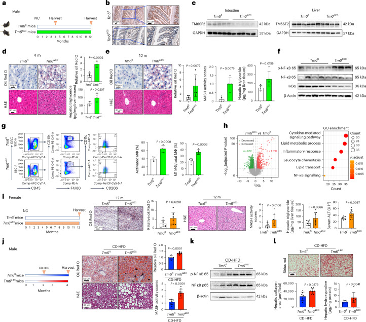
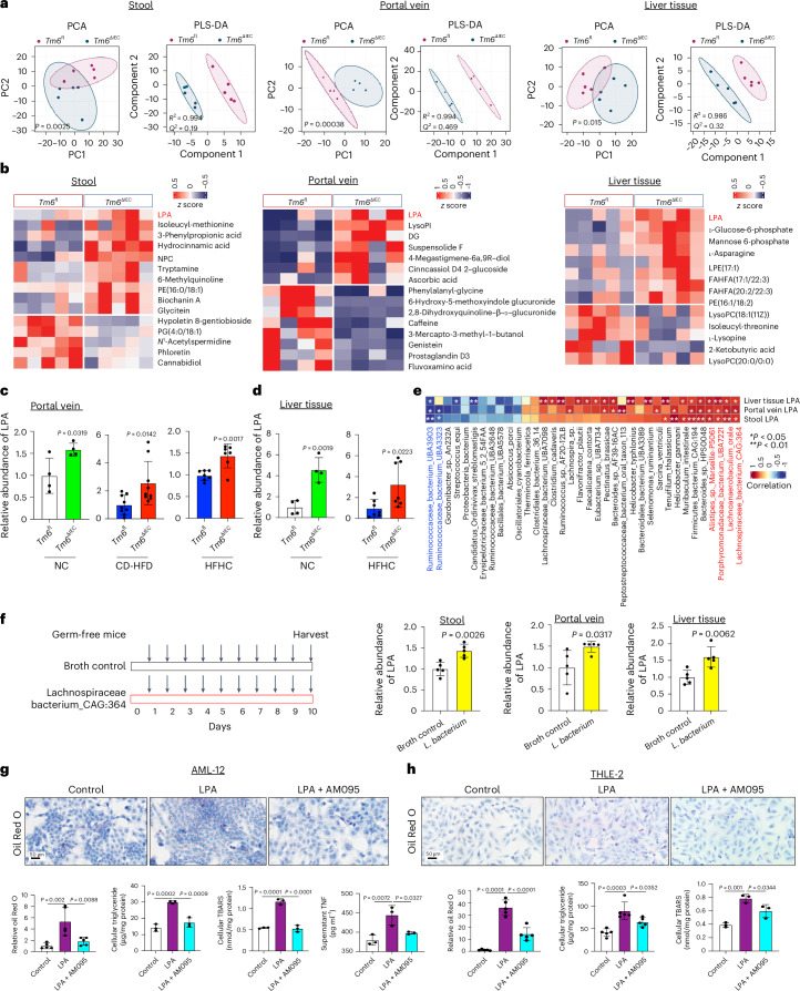
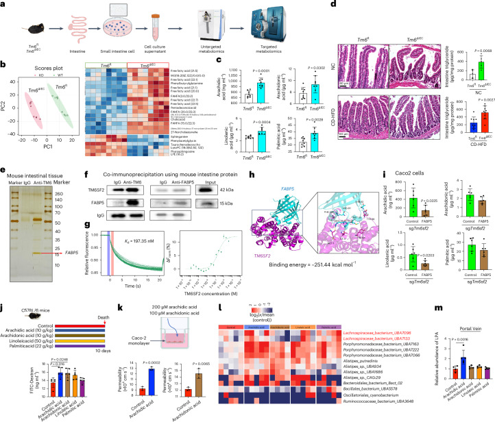
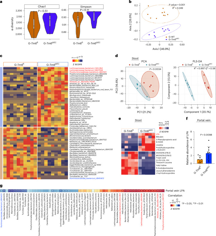
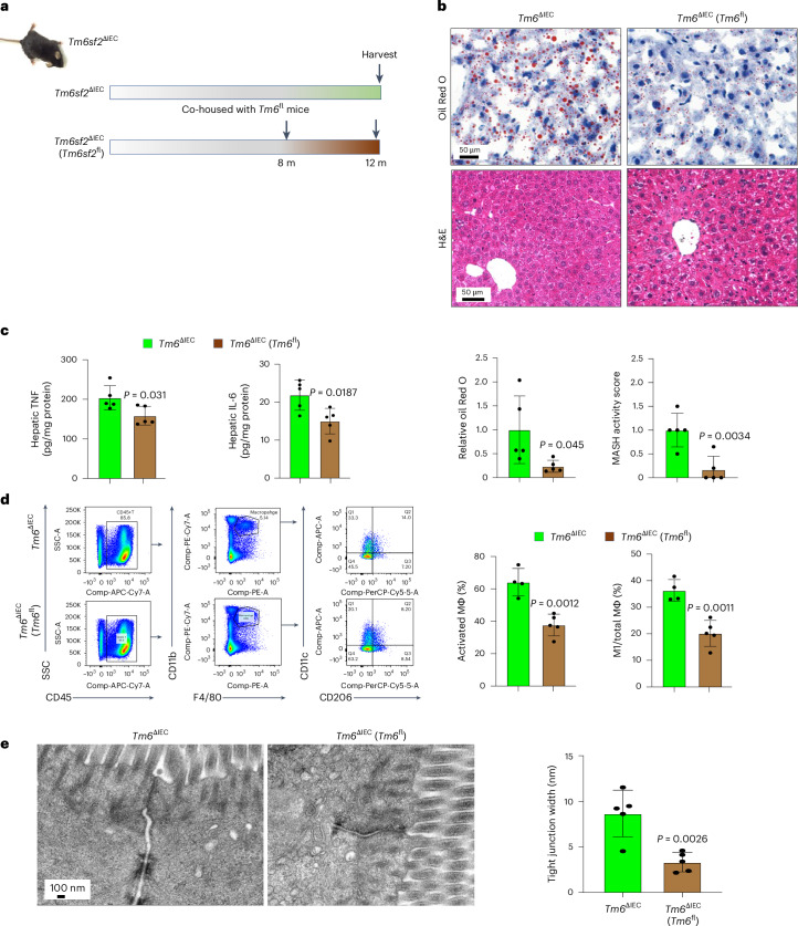
Transmembrane-6 superfamily member 2 (TM6SF2) regulates hepatic fat metabolism and is associated with metabolic dysfunction-associated steatohepatitis (MASH). TM6SF2 genetic variants are associated with steatotic liver disease. The pathogenesis of MASH involves genetic factors and gut microbiota alteration, yet the role of host-microbe interactions in MASH development remains unclear. Here, we discover that mice with intestinal epithelial cell-specific knockout of Tm6sf2 (Tm6sf2ΔIEC) develop MASH, accompanied by impaired intestinal barrier and microbial dysbiosis. Transplanting stools from Tm6sf2ΔIEC mice induces steatohepatitis in germ-free recipient mice, whereas MASH is alleviated in Tm6sf2ΔIEC mice co-housed with wild-type mice. Mechanistically, Tm6sf2-deficient intestinal cells secrete more free fatty acids by interacting with fatty acid-binding protein 5 to induce intestinal barrier dysfunction, enrichment of pathobionts, and elevation of lysophosphatidic acid (LPA) levels. LPA is translocated from the gut to the liver, contributing to lipid accumulation and inflammation. Pharmacological inhibition of the LPA receptor suppresses MASH in both Tm6sf2ΔIEC and wild-type mice. Hence, modulating microbiota or blocking the LPA receptor is a potential therapeutic strategy in TM6SF2 deficiency-induced MASH.


Conflict of interest statement

Competing interests: The authors declare no competing interests.

Figures

Fig. 1
Fig. 1. Intestinal Tm6sf2 deficiency in mice triggers MASH.
a, Experimental schematic of Tm6sf2ΔIEC and Tm6sf2fl male mice fed with NC for 4 or 12 months. b,c, Representative small intestine images of TM6SF2 immunohistochemistry (b), and TM6SF2 protein expression in small intestine and liver tissues (c) of Tm6sf2ΔIEC and Tm6sf2fl mice (n = 5 per group). d, Representative liver images of Oil Red O and H&E staining and hepatic triglyceride of Tm6sf2ΔIEC and Tm6sf2fl mice fed with NC for 4 months (n = 5 per group). eh, Representative liver images of Oil Red O and H&E staining with histological scoring and hepatic triglyceride (n = 5 per group; e) and hepatic protein expression of NF-κB pathway markers (Tm6sf2ΔIEC, n = 3; Tm6sf2fl, n = 5; f), flow cytometric analysis of hepatic macrophage (MΦ) populations (Tm6sf2ΔIEC, n = 4; Tm6sf2fl, n = 5; g) and volcano plot and Gene Ontology enrichment analysis of RNA sequencing on liver tissues (n = 5 per group; h) of Tm6sf2ΔIEC and Tm6sf2fl mice at 12 months of age. i, Experimental schematic, representative hepatic images of Oil Red O and H&E staining with histological scoring and hepatic triglyceride and serum ALT levels of Tm6sf2ΔIEC and Tm6sf2fl female mice fed with NC for 12 months (n = 9 per group). j,k, Experimental schematic and representative liver images of Oil Red O and H&E staining with histological scoring (n = 9 per group; j) and hepatic protein expression of NF-κB pathway markers (n = 3 per group; k) of Tm6sf2ΔIEC and Tm6sf2fl male mice fed with CD-HFD for 2 months. l, Representative liver images of Sirius Red staining and hepatic hydroxyproline of Tm6sf2ΔIEC and Tm6sf2fl mice fed with CD-HFD for 14 months (n = 7 per group). Results are presented as the mean ± s.d. Statistical significance was determined by two-tailed Student’s t-test (d, g, i and j), two-tailed Mann–Whitney U test (e and l), DESeq2 (h, left) or clusterProfiler (h, right). Tm6, Tm6sf2. Source data
Fig. 2
Fig. 2. Intestine Tm6sf2 deficiency results in intestinal barrier dysfunction and gut microbiota dysbiosis.
a,b, LPS level in portal vein serum (a) and liver tissues (b) of Tm6sf2ΔIEC and Tm6sf2fl mice fed with NC (n = 5 per group) or CD-HFD (n = 9 per group). c, LBP level in portal vein serum of Tm6sf2ΔIEC and Tm6sf2fl mice fed with CD-HFD (n = 9 per group). d, Transmission electron microscopy of Tm6sf2ΔIEC and Tm6sf2fl mice fed with NC at age 12 months (n = 5 per group) or CD-HFD for 2 months (n = 9 per group). e,f, Representative small intestine images of immunofluorescence staining for E-cadherin (red), villin (green) and DAPI (blue; e), and expression of intestinal barrier proteins in the small intestine (f) of NC-fed Tm6sf2ΔIEC and Tm6sf2fl mice (n = 5 per group) g, α-diversity (Chao1 or Simpson) and β-diversity between NC-fed Tm6sf2ΔIEC (n = 12) and Tm6sf2fl (n = 13) mice. The central horizontal line denotes the 50th percentile, while the box contains the 25th to 75th percentiles of the dataset. The whiskers mark the 5th and 95th percentiles. Data from two replicates were combined for analysis. h, Heat map of differential faecal microorganisms between Tm6sf2ΔIEC (n = 12) and Tm6sf2fl (n = 13) mice. Results are presented as the mean ± s.d. Statistical significance was determined by two-tailed Student’s t-test (a, right; b; c and d, right), two-tailed Mann–Whitney U test (a, left and d, left), Wilcoxon test (g, left) or Adonis test (g, right). Source data
Fig. 3
Fig. 3. Intestinal Tm6sf2 deficiency promotes MASH by inducing metabolite alteration.
a,b, PCA and PLS-DA of untargeted metabolomic profiling (a), and heat map of differential metabolites (b) in stools (n = 5 per group), portal vein serum (n = 4 per group) and liver tissues (n = 5 per group) of NC-fed Tm6sf2ΔIEC and Tm6sf2fl mice. c, LPA-targeted metabolomics on portal vein serum of Tm6sf2ΔIEC and Tm6sf2fl mice fed with NC (n = 4 per group), CD-HFD (n = 9 per group) or HFHC diet (n = 7 per group). d, LPA-targeted metabolomics on liver tissues of mice fed with NC (n = 4 per group) or HFHC diet (n = 7 per group). e, Correlation analysis between differential bacteria and LPA levels in stools, portal vein serum and liver tissues of Tm6sf2ΔIEC and Tm6sf2fl mice. *P < 0.05, **P < 0.01. f, LPS-targeted metabolomics in stools, portal vein serum and liver tissues of germ-free mice gavaged with Lachnospiraceae for 10 days (n = 5 per group). g,h, Representative images of Oil Red O staining with stained area normalized to cell number (n = 5 per group), cellular triglyceride and lipid peroxidation normalized to total protein content (n = 3 per group), and supernatant TNF level (n = 3 per group) of AML-12 mouse normal hepatocytes (g) or THLE-2 human normal hepatocytes (h) under LPA treatment with or without LPAR inhibitor AM095. Results are presented as the mean ± s.d. Statistical significance was determined by Adonis test (a), two-tailed Student’s t-test (c, d and f, left and right), two-tailed Mann–Whitney U test (f, middle), Spearman’s correlation analysis (e) or one-way analysis of variance (ANOVA) followed by Turkey’s multiple comparison (g and h). Source data
Fig. 4
Fig. 4. Intestinal cells with Tm6sf2 deficiency secrete free fatty acids to induce gut microbiota dysbiosis.
a, Experimental schematic of untargeted metabolomic profiling on the culture supernatant of IECs isolated from Tm6sf2ΔIEC and Tm6sf2fl mice. Created with BioRender.com. b, PCA analysis and heat map of differential metabolites secreted by IECs from mice (n = 8 per group). c, Metabolomics targeting free fatty acids on the cultural supernatant of isolated IECs (n = 8 per group). d, Representative small intestine images of H&E staining and intestinal triglyceride of Tm6sf2ΔIEC mice fed with NC (n = 5 per group) or CD-HFD (n = 9 per group). e, Silver staining coupled with mass spectrometry analysis after pull-down assay on mouse intestinal tissues. f, Co-immunoprecipitation of TM6SF2 and FABP5 using mouse intestinal proteins. g, MST assay for direct binding between TM6SF2 and FABP5 with dissociation constant (Kd) provided. h, Representative structure of molecular docking between TM6SF2 (purple) and FABP5 (blue). i, Metabolomics targeting free fatty acids in TM6SF2 KO Caco2 cells with or without FABP5 overexpression (n = 6 per group). j, Experimental schematic and intestinal permeability by FITC-labelled dextran assay in mice supplemented with the top differential free fatty acids (n = 5 per group). k, Permeability of Caco2 cell monolayers treated with arachidic acid or arachidonic acid (n = 3 per group). l,m, Heat map of differential faecal microorganisms (l) and LPA-targeting metabolomics on portal vein serum (m) of mice treated with free fatty acids (n = 4–5 per group). Results are presented as the mean ± s.d. Experiments were repeated three times with similar results (e and f). Statistical significance was determined by two-tailed Student’s t-test (c, d, i and k) or one-way ANOVA followed by Turkey’s multiple comparison (j and m). Source data
Fig. 5
Fig. 5. Gut microbiota from Tm6sf2ΔIEC mice promotes NASH development in germ-free mice.
a, Schematic of FMT experiment and representative images of Oil Red O and H&E staining with histological scoring in male germ-free mice transplanted with stools from Tm6sf2ΔIEC (G-Tm6sf2ΔIEC) or Tm6sf2fl (G-Tm6sf2fl) and fed with NC for 4 or 8 months (n = 10–11 per group). bd, Hepatic triglyceride and lipid peroxidation (b), visceral fat (c) and PCR array and Ccl1 and Ccl12 mRNA levels in liver tissues (d) of G-Tm6sf2ΔIEC and G-Tm6sf2fl mice 4 months after FMT (n = 10 per group). e, Representative images of Oil Red O and H&E staining with histological scoring in G-Tm6sf2ΔIEC (n = 11) and G-Tm6sf2fl (n = 10) 8 months after FMT. Results are presented as the mean ± s.d. Statistical significance was determined by two-tailed Student’s t-test (b and c) and two-tailed Mann–Whitney U test (a and e). TBARS, thiobarbituric acid-reactive substances assay. Source data
Fig. 6
Fig. 6. Gut microbiota and metabolomics in germ-free mice transplanted with stools from Tm6sf2ΔIEC mice.
a, α-diversity (Chao1 or Simpson) between G-Tm6sf2ΔIEC (n = 9) and G-Tm6sf2fl (n = 10) mice. The central horizontal line denotes the 50th percentile, while the box contains the 25th to 75th percentiles of the dataset. The whiskers mark the 5th and 95th percentiles. b,c, β-diversity (b) and heat map of differential faecal microorganisms (c) between G-Tm6sf2ΔIEC (n = 9) and G-Tm6sf2fl (n = 10) mice. d,e, PCA and PLS-DA analyses (d), and heat map of differential metabolites (e) identified by untargeted metabolomic profiling on stools of G-Tm6sf2ΔIEC and G-Tm6sf2fl mice (n = 5 per group). f, LPA-targeting metabolomics on portal vein serum of G-Tm6sf2ΔIEC and G-Tm6sf2fl mice (n = 10 per group). g, Correlation analysis between differential bacteria and portal vein LPA in G-Tm6sf2ΔIEC (n = 9) and G-Tm6sf2fl (n = 10) mice. *P < 0.05, **P < 0.01. Results are presented as the mean ± s.d. Statistical significance was determined by Wilcoxon test (a), Adonis test (b and d), two-tailed Student’s t-test (f) or Spearman’s rank correlation coefficient (g). P value was adjusted in g. Source data
Fig. 7
Fig. 7. Gut microbiota modulation improves MASH induced by intestinal Tm6sf2 deficiency.
a, Experimental schematic of microbiota modulation by co-housing male Tm6sf2∆IEC mice with male Tm6sf2fl mice for 4 months to restore the gut microbiota (n = 5 per group). be, Representative images of Oil Red O and H&E staining with histological scoring (b), hepatic levels of TNF and IL-6 (c), flow cytometric analysis of hepatic immune cell populations (d) and transmission electron microscopy (e) in Tm6sf2∆IEC and co-housed Tm6sf2∆IEC mice (n = 5 per group). Results are presented as the mean ± s.d. Statistical significance was determined by two-tailed Student’s t-test (be). Source data
Fig. 8
Fig. 8. Pharmacological inhibition of LPA receptor suppresses MASH.
a, Experimental schematic of 12-month-old male Tm6sf2∆IEC mice treated with LPAR inhibitor AM095 (10 mg per kg body weight) or vehicle control twice per week for 6 weeks. b,c, Representative images of Oil Red O and H&E staining with histological scoring (b), and hepatic triglyceride and hepatic lipid peroxidation (c) in Tm6sf2∆IEC mice treated with AM095 or vehicle control (n = 5 per group). df, Experimental schematic (d), representative images of Oil Red O and H&E staining with histological scoring (e), and hepatic triglyceride and serum ALT level (f) of male conventional C57BL/6 wild-type mice fed with NC (n = 5), MCD diet with vehicle control (n = 7), or MCD diet with LPAR inhibitor AM095 (n = 8) for 10 days. gi, Experimental schematic (g), representative images of Oil Red O and H&E staining with histological scoring (h) and hepatic triglyceride and lipid peroxidation (i) of male C57BL/6 wild-type mice fed with NC (n = 5), MCD diet with vehicle control (n = 5), or MCD diet with LPAR inhibitor Brp-LPA (n = 6). j, Overview schematic of the study. Created with BioRender.com. Results are presented as the mean ± s.d. Statistical significance was determined by two-tailed Student’s t-test (b and c) or one-way ANOVA followed by Turkey’s multiple comparison (e, f, h and i). DMSO, dimethylsulfoxide; i.p., intraperitoneal; TBARS, thiobarbituric acid-reactive substances assay. Source data
Extended Data Fig. 1
Extended Data Fig. 1. Systemic Tm6sf2 deficiency triggers MASH in mice.
(a) Experimental schematic of systematic Tm6sf2 KO and wildtype control mice fed with NC for 4 months; (b, c) Representative images of Tm6sf2 immunohistochemistry (b), and Tm6sf2 protein expression in liver tissues (c) of systematic Tm6sf2 KO and wildtype mice (n = 5 per group); (d-f) Representative images of Oil Red O and H&E staining with histological scoring (d), hepatic triglyceride and lipid peroxidation (e), and serum ALT level (f) in Tm6sf2 KO (n = 9) and wildtype mice (n = 5); (g-i) Experimental schematic and representative images of H&E staining with histological scoring (g), hepatic lipid peroxidation (h), and serum ALT level (i) of systematic Tm6sf2 KO and wildtype control mice fed with CD-HFD for 2 months (n = 8 per group). Results are presented as mean ± s.d. Statistical significance was determined by two-tailed Student’s t test (e, g-i) and Mann-Whitney U test (d and f). Source data
Extended Data Fig. 2
Extended Data Fig. 2. Liver Tm6sf2 deletion induces hepatic lipid accumulation in mice.
(a) Experimental schematic of Tm6sf2∆Liver and Tm6sf2fl mice fed with NC for 4 months; (b-e) Representative images of Tm6sf2 immunohistochemistry (b), hepatic Tm6sf2 protein expression (n = 5 per group) (c), representative images of Oil Red O and H&E staining with histological scoring (d), and hepatic triglyceride and lipid peroxidation (e) of Tm6sf2∆Liver (n = 12) and Tm6sf2fl mice (n = 8); (f-h) Experimental schematic and representative images of H&E staining with histological scoring (f), hepatic lipid peroxidation (g), and serum ALT level (h) of Tm6sf2∆Liver (n = 11) and Tm6sf2fl mice (n = 7) fed with CD-HFD for 2 months. Results are presented as mean ± s.d. Statistical significance was determined by two-tailed Student’s t test (e-h). Source data
Extended Data Fig. 3
Extended Data Fig. 3. Hepatic levels of pro-inflammatory cytokines in Tm6sf2ΔIEC mice.
(a, b) Hepatic TNF (a) and mRNA levels of pro-inflammatory cytokines (b) of NC-fed Tm6sf2∆IEC and Tm6sf2fl mice at 12-month-old (n=5 per group); (c) Hepatic TNF and IL-6 levels of CD-HFD-fed Tm6sf2ΔIEC and Tm6sf2fl mice (n = 9 per group). Results are presented as mean ± s.d. Statistical significance was determined by two-tailed Student’s t test (a-c). Source data
Extended Data Fig. 4
Extended Data Fig. 4. Intestine Tm6sf2 deficiency in mice accelerates HFHC diet-induced MASH.
(a) Experimental schematic of Tm6sf2∆IEC and Tm6sf2flo mice fed with HFHC diet; (b) Representative hepatic images of H&E staining with histological scoring of HFHC-fed Tm6sf2∆IEC and Tm6sf2flo mice (n = 7 per group); (c, d) Representative small intestine images of transmission electron microscopy with tight junction width (c), and H&E staining (d) of HFHC-fed Tm6sf2ΔIEC and Tm6sf2flox mice (n = 7 per group). Results are presented as mean ± s.d. Statistical significance was determined by two-tailed Student’s t test (b) and two-tailed Mann-Whitney U test (c). Source data
Extended Data Fig. 5
Extended Data Fig. 5. Tm6sf2 knockout increases intestinal epithelial cell permeability in vitro.
Permeability of Caco2 cell monolayers treated with sgRNA-Tm6sf2 (n = 3 per group). Results are presented as mean ± s.d. Statistical significance was determined by one-way ANOVA followed by Turkey's multiple comparison. Source data
Extended Data Fig. 6
Extended Data Fig. 6. Functional prediction of LPA-producing bacteria.
(a) NAD(P)H-dependent glycerol-3-phosphate dehydrogenase [1.1.1.94] (gene: gpsA) and glycerol-3-phosphate 1-O-acyltransferase PlsY [2.3.1.15] (gene: plsY) are the key enzymes catalyzing glycerone phosphate to sn-glycerol 3-phosphate, the precursor of LPA; (b) Reference genomes of P. bacterium with genes encoding gpsA, and L. bacterium and A. marseille with genes encoding both plsY and gpsA.
Extended Data Fig. 7
Extended Data Fig. 7. Western blot analysis of NPC1L1 and CD36.
NPC1L1 and CD36 protein expression in the small intestine of Tm6sf2∆IEC andTm6sf2flox mice (n = 5 per group). Housekeeping controls are identical to the left panel of Fig. 1c.
Extended Data Fig. 8
Extended Data Fig. 8. Gut microbiota modulation by co-housing.
(a, b) PCA analysis (a) and heatmap of differential fecal microbes (b) of Tm6sf2∆IEC (n = 5) and co-housed Tm6sf2∆IEC mice (n = 3). Statistical significance was determined by adonis (a). Source data
Extended Data Fig. 9
Extended Data Fig. 9. LPAR inhibitor impairs HFHC diet-induced lipid accumulation and liver injury in mice.
(a-c) Experimental schematic (a), hepatic triglyceride (b), and serum ALT level (c) of C57BL/6 wildtype mice fed with HFHC diet for 3 months and treated with vehicle control (n = 9) or LPAR inhibitor AM095 (n = 10) for 1 month. Results are presented as mean ± s.d. Statistical significance was determined by two-sided Mann-Whitney U test (b, c). Source data
Extended Data Fig. 10
Extended Data Fig. 10. Strategies for generating conditional Tm6sf2 KO mice and systematic Tm6sf2 KO mice.
(a) Conditional Tm6sf2 KO C57BL/6 mice were generated through targeted deletion of exons 2-5, induced by Cre-loxP recombination in tissue-specific manner; (b) Systematic Tm6sf2 KO C57BL/6 mice were generated using CRISPR/Cas9 system by deleting a 4-base pair in the first exon of Tm6sf2 gene.

References

    1. Loomba, R., Friedman, S. L. & Shulman, G. I. Mechanisms and disease consequences of nonalcoholic fatty liver disease. Cell184, 2537–2564 (2021). - PubMed
    1. Yu, J., Shen, J., Sun, T. T., Zhang, X. & Wong, N. Obesity, insulin resistance, NASH and hepatocellular carcinoma. Semin. Cancer Biol.23, 483–491 (2013). - PubMed
    1. Eslam, M., Valenti, L. & Romeo, S. Genetics and epigenetics of NAFLD and NASH: clinical impact. J. Hepatol.68, 268–279 (2018). - PubMed
    1. Kozlitina, J. et al. Exome-wide association study identifies a TM6SF2 variant that confers susceptibility to nonalcoholic fatty liver disease. Nat. Genet.46, 352–356 (2014). - PMC - PubMed
    1. Mahdessian, H. et al. TM6SF2 is a regulator of liver fat metabolism influencing triglyceride secretion and hepatic lipid droplet content. Proc. Natl Acad. Sci. USA111, 8913–8918 (2014). - PMC - PubMed

Substances

LinkOut - more resources