. 2025 Feb;27(2):202-217.
doi: 10.1038/s41556-024-01580-y. Epub 2025 Jan 8.

H3K36 methylation regulates cell plasticity and regeneration in the intestinal epithelium

Affiliations

H3K36 methylation regulates cell plasticity and regeneration in the intestinal epithelium

Alison R S Pashos et al. Nat Cell Biol. 2025 Feb.

Abstract

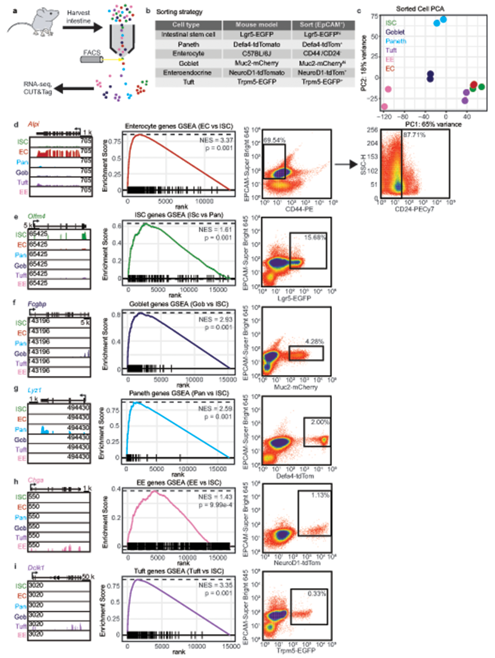
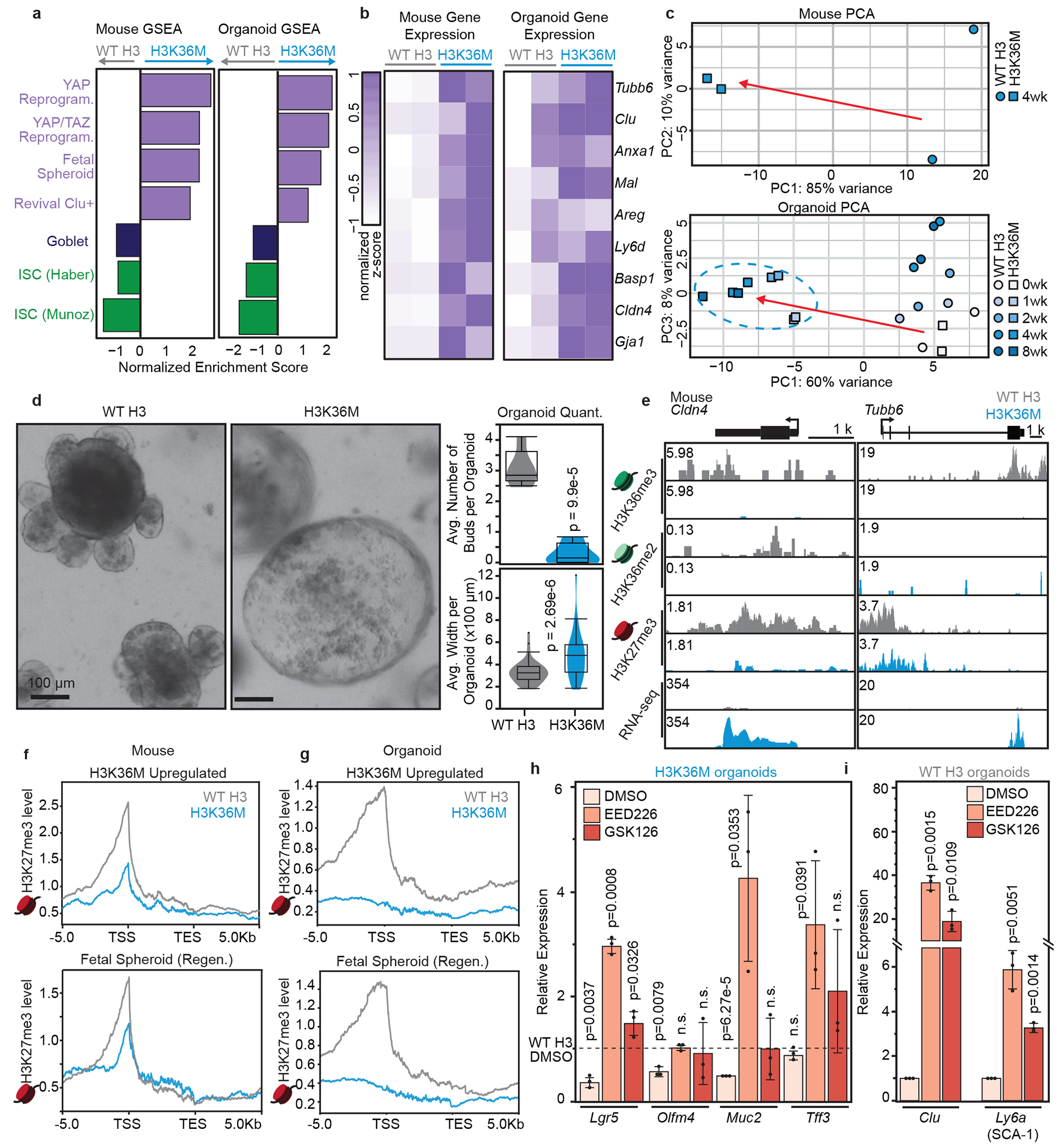
Plasticity is needed during development and homeostasis to generate diverse cell types from stem and progenitor cells. Following differentiation, plasticity must be restricted in specialized cells to maintain tissue integrity and function. For this reason, specialized cell identity is stable under homeostatic conditions; however, cells in some tissues regain plasticity during injury-induced regeneration. While precise gene expression controls these processes, the regulatory mechanisms that restrict or promote cell plasticity are poorly understood. Here we use the mouse small intestine as a model system to study cell plasticity. We find that H3K36 methylation reinforces expression of cell-type-associated genes to maintain specialized cell identity in intestinal epithelial cells. Depleting H3K36 methylation disrupts lineage commitment and activates regenerative gene expression. Correspondingly, we observe rapid and reversible remodelling of H3K36 methylation following injury-induced regeneration. These data suggest a fundamental role for H3K36 methylation in reinforcing specialized lineages and regulating cell plasticity and regeneration.


Conflict of interest statement

Competing interests: B.D.S. is a cofounder of EpiCypher. The other authors declare no competing interests.

Figures

Extended Data Fig. 1.
Extended Data Fig. 1.. Intestinal epithelial cells have distinct gene expression profiles.
(a) A schematic of the strategy used to sort and profile intestinal epithelial cell types. (b) Mouse lines used to sort intestinal epithelial cell types. (c) Multidimensional scaling analysis based on RNA-seq from sorted intestinal epithelial cells (n = 2 biological replicates each). (d-i) Gene tracks showing RNA-seq reads at representative genes (left), gene set enrichment analysis based on RNA-seq data for each sorted cell type using published gene sets (middle), and FACS plots (right) showing sorted cell populations (black box) for enterocytes (d), ISCs (e), goblet cells (f), Paneth cells (g), enteroendocrine cells (h), and tuft cells (i). Percentages in FACS plots represent percentage of live cells in each gate. Statistics were generated in accordance with the FGSEA algorithm(ref. 99).
Extended Data Fig. 2.
Extended Data Fig. 2.. Intestinal epithelial cells have distinct enrichment of GO terms.
(a) Top 10 GO terms enriched in goblet, Paneth, tuft, enteroendocrine, and enterocyte cells, compared to ISCs, based on differential gene expression. Statistics were generated in accordance with the clusterProfiler algorithm for GO analysis. (b) Top 10 GO terms enriched in goblet, Paneth, tuft, enteroendocrine, and enterocyte cells, compared to ISCs, based on differential H3K36me3. Statistics were generated in accordance with the clusterProfiler algorithm for GO analysis.
Extended Data Fig. 3.
Extended Data Fig. 3.. Intestinal epithelial cells have differential H3K36me3 enrichment.
(a) Meta-analysis of H3K36me3 at published cell type-associated genes in each sorted cell type (5 kb window around the TSS and TES, representative trace for 2 biological replicates). (b) Correlation analysis for H3K36me3 and H3K27me3 in 10,000 randomly selected bins across the genome. The color of each dot represents gene expression (counts) in that bin. (c,e,g,i) Gene tracks for H3K36me3 at Dclk1, a tuft cell-associated gene (c), Alpi, an enterocyte-associated gene (e), Chga, an enteroendocrine cell-associated gene (g), and Lgr5, an ISC-associated gene (i) (representative track for 2 biological replicates). (d,f,h) Left, boxplots for H3K36me3 levels for a published tuft cell-associated gene set (d), enterocyte-associated gene set (f), or enteroendocrine-associated gene set (h) and all other expressed genes; right, boxplots for gene expression for a published tuft cell-associated gene set (d), enterocyte-associated gene set (f), or enteroendocrine-associated gene set (h) and all other expressed genes (n = 2 biological replicates; box plot shows the median, box plot center lines represent the median, box edges represent the first and third quartiles, and whiskers indicate minimum and maximum values). (j) Representative gene tracks for H3K36me3 and RNA-seq data for each sorted cell type over Gfi1, a Paneth- and goblet-specific gene (n = 2 biological replicates). (k) Representative gene tracks of H3K36me3 and RNA-seq data for each sorted cell type over Mettl1 and Commd4, non-cell type-specific genes that are expressed in Paneth and goblet cells (n = 2 biological replicates).
Extended Data Fig. 4.
Extended Data Fig. 4.. H3K36me3 is largely distinct between intestinal epithelial cell types.
(a) Pairwise comparison of H3K36me3 between the indicated cell types. Colored dots indicate peaks over genes differentially expressed between each cell type. Gray dots represent 10,000 randomly selected peaks over non-significantly differentially expressed genes. (b) Pairwise comparison of H3K27me3 between the indicated cell types. Colored dots indicate peaks over genes differentially expressed between each cell type. Gray dots represent 10,000 randomly selected peaks over non-significantly differentially expressed genes.
Extended Data Fig. 5.
Extended Data Fig. 5.. Suppressing H3K36 methylation disrupts intestinal homeostasis.
(a) Western blot analysis of β-ACTIN, H3K36M, and total H3 levels in C57Bl6, WT H3 no dox, H3K36M no dox, WT H3 dox, and H3K36M dox mice. (b) Representative images of immunofluorescence for H3 in C57Bl6, WT H3 no dox, H3K36M no dox, WT H3 dox, and H3K36M dox mice. Scale bar = 50 μm. (c) Periodic acid-Schiff stain on intestinal sections from WT H3 and H3K36M mice treated for 4 weeks with dox. Black arrows indicate secretory cells (n = 2 mice each). Scale bar = 50 μm. (d) Representative images of immunofluorescence for CHGA, a marker of enteroendocrine cells, in WT H3 and H3K36M intestinal sections. The inset is a magnified image of a positive cell. Scale bar=50 μm. (left, n = 2 mice each genotype). Quantification of enteroendocrine cells (right, n=10 images per mouse; unpaired two-tailed student’s t-test; box plot center lines represent the median, box edges represent the first and third quartiles, and whiskers indicate minimum and maximum values). (e) Representative images of immunofluorescence for DCLK1, a marker of tuft cells, in WT H3 and H3K36M intestinal sections. The inset is a magnified image of a positive cell. Scale bar=50 μm. (left, n = 2 mice each genotype). Quantification of enteroendocrine cells (right, n = 10 images per mouse; unpaired two-tailed student’s t-test; box plot center lines represent the median, box edges represent the first and third quartiles, and whiskers indicate minimum and maximum values). (f) Representative transmission electron microscopy images of a WT H3 enterocyte cell (top, scale bar 2 μm), and H3K36M enterocyte cell found in the villus (bottom, scale bar 2 μm). The inset shows the brush border in each respective cell type (n = 3 mice each). (g) Representative images of immunofluorescence for γ-ACTIN, E-CADHERIN, and DAPI on WT H3 and H3K36M intestine sections after four weeks dox induction (left; n = 2 mice each) and quantification (right; n = 2 mice each). Scale bar = 2 μm.
Extended Data Figure 6.
Extended Data Figure 6.. Expressing H3K36M disrupts chromatin and gene expression at cell type-associated genes.
(a) H3K36me3 signal over all genes in representative WT H3 and H3K36M samples (n = 2 biological replicates each). (b) H3K36me2 levels at all domains of H3K36me2 in representative WT H3 and H3K36M samples (n = 2 biological replicates each). (c) Representative gene tracks for H3K36me3, H3K36me2, H3K27me3, and RNA-seq for WT H3 and H3K36M samples over Otop3 and Dpp4, markers of enterocytes (n = 2 biological replicates each). (d) Representative gene tracks for H3K36me3, H3K36me2, H3K27me3, and RNA-seq for WT H3 and H3K36M samples over Fcgbp, a marker of goblet cells, and Olfm4 and Lgr5, markers of ISCs (n = 2 biological replicates each). (e) Representative gene tracks for H3K36me3 and RNA-seq over Mettl1 and Commd4, non-cell type-specific genes, and Gfi1, a goblet/ Paneth cell marker (n = 2 biological replicates each). (f) Representative gene tracks for RNA-seq over PRC2 components Eed, Ezh2, Suz12, Aebp2, Phf1, and Rbbp4 in WT H3 and H3K36M mice (n = 2 biological replicates each). (g) Representative gene tracks for H3K36me2 and H3K27me3 over genic (white) and intergenic (orange) regions in WT H3 and H3K36M samples (n = 2 biological replicates each). (h) Quantification of H3K36me2 counts in 10,000 randomly selected 10 kb bins from intergenic regions across the mouse genome in WT H3 and H3K36M mice (Wilcoxon test; box plot center lines represent the median, box edges represent the first and third quartiles, and whiskers indicate minimum and maximum values). (i) Quantification of H3K27me3 counts in 10,000 randomly selected 10 kb bins from intergenic regions across the mouse genome in WT H3 and H3K36M mice (Wilcoxon test; box plot center lines represent the median, box edges represent the first and third quartiles, and whiskers indicate minimum and maximum values). (j) Quantification of H3K36me3 binned into 10 kb regions over the gene body of immature Paneth genes, mature Paneth population 1 genes, and mature Paneth population 2 genes from Fig. 4 (Wilcoxon test; box plot center lines represent the median, box edges represent the first and third quartiles, and whiskers indicate minimum and maximum values). (k) Quantification of H3K27me3 binned into 10 kb regions over the gene body of immature Paneth genes, mature Paneth population 1 genes, and mature Paneth population 2 genes from Fig. 4 (Wilcoxon test; box plot center lines represent the median, box edges represent the first and third quartiles, and whiskers indicate minimum and maximum values). (l) Quantification of H3K36me2 binned into 5 kb regions upstream of immature Paneth genes, mature Paneth population 1 genes, and mature Paneth population 2 genes from Fig. 4 (Wilcoxon test; box plot center lines represent the median, box edges represent the first and third quartiles, and whiskers indicate minimum and maximum values). (m) Quantification of H3K27me3 binned into 5 kb regions supstream of immature Paneth genes, mature Paneth population 1 genes, and mature Paneth population 2 genes from Fig. 4 (Wilcoxon test; box plot center lines represent the median, box edges represent the first and third quartiles, and whiskers indicate minimum and maximum values).
Extended Data Fig. 7.
Extended Data Fig. 7.. Intestinal epithelium specific H3K36M expression recapitulates phenotypes observed in whole body H3K36M mice.
(a) A schematic of design of V5-WT H3 and V5-H3K36M mice (top), and of the experimental design for induction of these mice (bottom). (b) Western blot analysis of bulk intestinal epithelium. (c) Representative images of immunofluorescence in V5-WT H3 and V5-H3K36M intestines (n = 2 mice each). The inset is a magnified image of the epithelium. Scale bar = 50 μm. (d) H&E stain on intestinal sections from V5-WT H3 and V5-H3K36M mice at 4 weeks. Black arrows indicate secretory cells (n = 2 mice each). Scale bar = 50 μm. (e) Representative images of immunofluorescence for LYZ1 (magenta), TFF3 (green), and DAPI (blue) in V5-WT H3 and V5-H3K36M intestines (n =2 mice each). The inset is a magnified image of a double positive cell. Scale bar = 50 μm. (f) qRT-PCR on V5-WT H3 and V5-H3K36M mice for a marker of stem cells (Olfm4), Paneth cells (Lyz1), and secretory cells (Muc2) (n = 3 mice; unpaired two-tailed student’s t-test; data are presented as mean values +/−standard deviation). (g,h) Representative images of immunofluorescence for OLFM4 (g) and KI67 and EdU after 24h chase (h) on V5-WT H3 and V5-H3K36M mice at 8 weeks (left; n = 4 mice each) and quantification (right; n = 10 images per mouse; unpaired two-tailed student’s t-test). (i,k) Normalized enrichment scores from GSEA based on RNA-seq comparing WT H3 and H3K36M mice after four weeks of induction, using gene sets for DNA damage response (i) and inflammation (k). (j,l) Representative images of immunofluorescence for γH2A-X, a mark of DNA damage (j), and CD45, a mark of inflammatory cells (l), on V5-WT H3 and V5-H3K36M mice at 2, 3, and 4 weeks (left, n = 3 mice each) and quantification (right; n = 10 images per mouse; unpaired two-tailed student’s t-test). (m) Weight per week of each V5-WT H3 and V5-H3K36M mouse (n = 4 mice). For all box plots, center lines represent the median, box edges represent the first and third quartiles, and whiskers indicate minimum and maximum values.
Extended Data Fig. 8.
Extended Data Fig. 8.. Expressing H3K36M in intestinal organoids recapitulate phenotypes observed in mice.
(a) Schematic of organoid derivation and treatment. (b) Western blot analysis at one week (n = 2 biological replicates). (c) Representative images of immunofluorescence (n = 2 biological replicates). Scale bar = 50 μm. (d) Normalized enrichment scores from GSEA based on RNA-seq at two weeks. (e) Representative images of immunofluorescence staining at eight weeks. Scale bar = 100 μm. (n = 2 biological replicates). (f) Differential peaks at four weeks. Yellow dots indicate peaks over significantly downregulated genes from RNA-seq (n=2 biological replicates; downregulated genes have p-value < 0.05 and log2FC > 1). (g) Gene expression in mice vs. organoids at 4 weeks. Yellow dots correspond to yellow dots in Fig. 3a and Extended Data Fig. 8f and are quantified in each quadrant. (h) Quantification of H3K36me3. (i,j,k,l) Quantification of H3K36me3 (i) and H3K27me3 (j) binned into 10 kb regions over the gene body, and H3K36me2 (k) and H3K27me3 (l) binned into 5 kb regions upstream of the gene body. (m,n,o,p) Quantification of H3K36me3 (m) and H3K27me3 (n) binned into 10 kb regions over the gene body, and H3K36me2 (o) and H3K27me3 (p) binned into 5 kb regions upstream of the gene body. (q,r) GSEA based on RNA-seq of mice (q) and organoids (r) (plots correspond to NES in Fig. 5a, left (q) and right (r)). Statistics were generated in accordance with the FGSEA algorithm. (s) qRT-PCR for markers of regeneration (n = 3 or 4 mice as indicated; unpaired two-tailed student’s t-test; data are presented as mean values +/− standard deviation; p value indicates significance compared to V5-WT H3 at the same timepoint and gene). (t) Gene expression in organoids. Each time point is normalized to day 0. Notable regenerative markers are labeled. (u) Representative gene tracks in organoids over Cldn4 and Tubb6 (n = 2 biological replicates). (v) Western blot analysis in organoids. For all box plots, center lines represent the median, box edges represent the first and third quartiles, and whiskers indicate minimum and maximum values.
Extended Data Fig. 9.
Extended Data Fig. 9.. Knockdown of H3K36 methyltransferases, Nsd2 or Setd2, phenocopy H3K36M expression in organoids.
(a,b) Western blot analysis for organoids transduced with shRNAs. (c,d) qRT-PCR on shNsd2 (c) or shSetd2 (d) organoids for markers of stem cells, secretory cells, or regeneration (n = 3 independent replicates per shRNA; unpaired two-tailed student’s t-test; data is represented as mean values +/− standard deviation). (e) Representative images of immunofluorescence for MMP7, a marker of Paneth cells, and MUC2, a marker of goblet cells. Scale bar = 100 μm. (n = 2 replicates per target). (f,g) Left, representative brightfield images of shNsd2 (f) and shSetd2 (g) organoids. Scale bar = 100 μm. Middle, quantification of organoid budding in shNsd2 (f) and shSetd2 (g) organoids (middle, n = 10 images each; unpaired two-tailed student’s t-test). Quantification of organoid width in shNsd2 (f) and shSetd2 (g) organoids. (right, n=10 images each; unpaired two-tailed student’s t-test). Box plot center lines represent the median, box edges represent the first and third quartiles, and whiskers indicate minimum and maximum values. (h,i,j) Metaplot and heatmaps of H3K27me3 and EZH2 (h), H3K36me2 and NSD2 (i), H3K36me3 and SETD2 (j) signal in representative WT H3 and H3K36M samples (n = 2 biological replicates each). (k,l) Representative gene tracks for H3K27me3 and EZH2 over Lgr5 and Olfm4, markers of ISCs (k), and Tubb6 and Cldn4, markers of regeneration (l, n = 2 biological replicates each). (m,n) Representative gene tracks for H3K36me2 and NSD2 over Lgr5 and Olfm4, markers of ISCs (m), and Tubb6 and Cldn4, markers of regeneration (n, n = 2 biological replicates each). (o,p) Representative gene tracks for H3K36me3 and SETD2 over Lgr5 and Olfm4, markers of ISCs (o), and Tubb6 and Cldn4, markers of regeneration (p, n = 2 biological replicates each).
Extended Data Fig. 10.
Extended Data Fig. 10.. H3K36 methylation is remodeled following injury-induced regeneration in organoids.
(a) Weight per day after 12 Gy irradiation of each mouse over 5 days (n = 6 mice). (b) Differential methylation analysis between WT non-irradiated and 24-hour post-irradiation (left column), 48-hour post-irradiation (middle column), and 96-hour post-irradiation (right column) organoids in 10kb bins across the genome. In the top row, purple dots indicate bins containing published fetal spheroid-associated genes. In the bottom row, green dots represent ISC-associated genes. Up to 5000 random datapoints are plotted at each timepoint. Percentages in each quadrant represent the percentage of total colored dots in the graph that fall in that quadrant. (c) Differential methylation analysis as in panel b, but with gene expression values overlaid on only colored dots from panel b (log2FC gene expression in that bin). (d) Representative images of WT H3 and H3K36M organoids 7 days after irradiation and single cell plating. Right images are insets of left images. Scale bar = 100 μm. (e) A schematic representation of chromatin in the intestine under homeostatic conditions. (f) Upon H3K36M induction or injury, H3K36me2 and H3K36me3 are depleted across the genome. H3K27me3 spreads into intergenic regions and cell-type associated genes and is titrated away from regenerative genes. Changes to the chromatin landscape return to normal, homeostatic levels as regeneration completes.
Fig. 1.
Fig. 1.. Intestinal epithelial cells have distinct H3K36 methylation profiles at cell type-associated genes.
(a) A schematic of a typical crypt-villus intestinal unit. (b) A simplified schematic of differentiation in the intestinal epithelium. (c) Meta-analysis of H3K36me3 over published cell type-associated genes in each sorted cell type (5 kb window around the transcription start site (TSS) and transcription end site (TES), representative trace for 2 biological replicates). The goblet cell and Paneth cell gene sets are modified from Haber et al. (see methods for details). Dark green nucleosome icons represent H3K36me3 throughout the paper. (d) Differential H3K36me3 peaks between ISCs and Paneth cells. Colored dots indicate peaks over differentially expressed genes (blue = upregulated genes in Paneth cells, green = upregulated genes in ISCs) and the number of differentially expressed genes with differential H3K36me3 is indicated. (e) Differential H3K27me3 peaks between ISCs and Paneth cells. Colored dots indicate peaks over differentially expressed genes (blue = upregulated genes in Paneth cells, green = upregulated genes in ISCs) and the number of differentially expressed genes with differential H3K27me3 is indicated. Dark red nucleosome icons represent H3K27me3 throughout the paper. (f,h,j) Gene tracks for H3K36me3 at Olfm4, an ISC-associated gene (f), Fcgbp, a goblet cell-associated gene (h), and Lyz1, a Paneth cell-associated gene (j) (representative track for 2 biological replicates). (g,i,k) Left, boxplots for H3K36me3 levels for a published ISC-associated gene set (g), goblet cell-associated gene set (i), or Paneth cell-associated gene set (k), and all other expressed genes; right, boxplots for gene expression for a published ISC-associated gene set (g), goblet cell-associated gene set (i), or Paneth cell-associated gene set (k), and all other expressed genes (n = 2 biological replicates, box plot center lines represent the median, box edges represent the first and third quartiles, and whiskers indicate minimum and maximum values).
Fig. 2.
Fig. 2.. Suppressing H3K36 methylation disrupts adult intestinal homeostasis.
(a) A schematic of the experimental design for dox induction and harvest of WT H3 and H3K36M intestinal epithelium. (b) Western blot analysis of bulk intestinal epithelium. (c) Representative images of immunofluorescence in WT H3 and H3K36M intestines (n = 3 WT H3 mice, 2 H3K36M mice). The inset is a magnified image of the epithelium. Light green nucleosome icons represent H3K36me2 throughout the paper. (d) Quantification of immunofluorescence (n = 5 mice each genotype, 10 images per mouse; unpaired two-tailed student’s t-test; box plot center lines represent the median, box edges represent the first and third quartiles, and whiskers indicate minimum and maximum values). (e,f,g,h) Representative images of immunofluorescence for OLFM4 (e), KI67 (f), MUC2 (g), or LYZ1 (h) on WT H3 and H3K36M intestine sections after four weeks dox induction (left; n = 5 mice each) and quantification (right; n = 10 images per mouse; unpaired two-tailed student’s t-test; box plot center lines represent the median, box edges represent the first and third quartiles, and whiskers indicate minimum and maximum values).
Fig. 3.
Fig. 3.. Expressing H3K36M disrupts chromatin and gene expression at cell type-associated genes.
(a) Differential peaks for H3K36me3 between WT H3 and H3K36M mice induced for four weeks. Yellow dots indicate peaks occurring over significantly downregulated genes in H3K36M from RNA-seq (n = 2 mice each; downregulated genes defined as p-value < 0.05 and log2FC > 1). (b) Quantification of H3K36me3 for all peaks over genes, H3K36M downregulated genes, or H3K36M upregulated genes (box plot center lines represent the median, box edges represent the first and third quartiles, and whiskers indicate minimum and maximum values). (c) Normalized enrichment scores from gene set enrichment analysis based on RNA-seq from WT H3 and H3K36M intestinal epithelial cells after four weeks dox induction using published cell type-associated gene sets. (d,e) Meta-analysis of H3K36me3 (d) and H3K27me3 (e) over genes downregulated H3K36M samples (5 kb window around the TSS and TES, representative trace from two biological replicates). (f,g) Quantification of H3K36me3 (f) and H3K27me3 (g), binned into 10 kb regions over the gene body of genes downregulated in H3K36M samples, ISC-associated genes, and goblet cell-associated genes (box plot center lines represent the median, box edges represent the first and third quartiles, and whiskers indicate minimum and maximum values). (h,i) Meta-analysis of H3K36me2 (h) or H3K27me3 (i) over genes downregulated H3K36M samples (25 kb window around the TSS and TES, representative trace from two biological replicates). (j,k) Quantification of H3K36me2 (j) or H3K27me3 (k), binned into 5 kb regions upstream of the gene body of genes downregulated in H3K36M samples, ISC-associated genes, and goblet cell-associated genes (box plot center lines represent the median, box edges represent the first and third quartiles, and whiskers indicate minimum and maximum values). (l,m) Meta-analysis of H3K27me3 over ISC-associated genes (l) or goblet-associated genes (m) (25 kb window around the TSS and TES, representative trace from two biological replicates).
Fig. 4.
Fig. 4.. Suppressing H3K36 methylation induces the accumulation of abnormal secretory cells.
(a) Left, representative immunofluorescence images for LYZ1 and TFF3 on WT H3 and H3K36M intestine sections after four weeks dox induction (n = 5 mice each, white arrows indicate LYZ1+/TFF3+ cells). Right, quantification of LYZ1+/TFF3+ cells (n = 10 images per mouse; unpaired two-tailed student’s t-test; box plot center lines represent the median, box edges represent the first and third quartiles, and whiskers indicate minimum and maximum values). (b) Representative transmission electron microscopy images of a WT H3 goblet cell (left, scale bar 2 μm), WT H3 Paneth cell (middle, scale bar 2 μm) and H3K36M intermediate cell found in the villus (right, scale bar 500 nm). The inset shows granules in each respective cell type (n = 3 mice each). (c) UMAP representation for unsupervised clustering of 3 distinct Paneth cell clusters sorted from Defa4Cre; Rosa26tdTomato mice. (d) UMAP representation from panel c overlaid with expression values for the mature Paneth cell gene Defa20 and immature Paneth cell gene Mmp7. Dark purple indicates maximum gene expression, while light purple indicated low or no expression in log-normalized unique molecular identifier counts. (e) Expression level and percent of cells in each cluster expressing the indicated genes. (f) Normalized enrichment scores from gene set enrichment analysis based on RNA-seq from WT H3 and H3K36M intestines after four weeks dox induction using gene sets established from Paneth cell scRNA-seq clusters.
Fig. 5.
Fig. 5.. Suppressing H3K36 methylation activates a regenerative gene expression signature.
(a) Gene set enrichment analysis based on RNA-seq from WT H3 and H3K36M mice (left) and WT H3 and H3K36M organoids (right) after four weeks dox induction, using published gene sets related to regeneration (purple) and mature intestinal epithelial cells. (b) Gene expression normalized z-score from WT H3 or H3K36M mice (left) and WT H3 and H3K36M organoids (right) for the indicated regenerative genes. (c) Principal component analysis from RNA-seq for WT H3 and H3K36M mice at 4 weeks of dox induction (top) and for WT H3 and H3K36M organoids over an 8-week time course (bottom, n = 2 biological replicates per condition). (d) Left, representative brightfield images of WT H3 and H3K36M intestinal organoids after 8 weeks of doxycycline induction (n = 3 independent replicates of 2 biological replicates each; scale bar 100 μm). Right, quantification of organoid budding and width in WT H3 and H3K36M organoids (n=10 images each; unpaired two-tailed student’s t-test). (e) Representative gene tracks for H3K36me3, H3K36me2, H3K27me3, and gene expression for Cldn4 and Tubb6, regenerative markers, in WT H3 and H3K36M mice. (f,g) Meta-analysis of H3K27me3 at genes upregulated in H3K36M mice (f) or organoids (g) (top) and at a published set of regenerative genes (bottom) (5 kb window around the TSS and TES, representative trace from two biological replicates). (h) qRT-PCR on H3K36M organoids treated with DMSO, EED226, or GSK126 for markers of stem cells (Lgr5, Olfm4) and secretory cells (Muc2, Tff3) (n = 3 independent replicates per treatment; unpaired two-tailed student’s t-test; data are presented as mean values with errors bars to represent standard deviation; p value indicates significance with regard to WT H3 DMSO for each gene). (i) qRT-PCR on WT H3 organoids treated with DMSO, EED226, or GSK126 for markers of regeneration (Clu, Ly6a) (n = 3 independent replicates per treatment; unpaired two-tailed student’s t-test; data are presented as mean values with errors bars to represent standard deviation; p value indicates significance with regard to WT H3 DMSO for each gene).
Fig. 6.
Fig. 6.. H3K36 methylation is remodeled following injury-induced regeneration in mice.
(a) A schematic of experimental design for irradiation of intestinal epithelial cells. (b) Normalized gene expression counts based on RNA-seq at each timepoint in irradiated mice for Clu, a marker of regeneration, and Lgr5, a marker of ISCs (n = 2 biological replicates). (c) Gene set enrichment analysis based on RNA-seq data comparing WT non-irradiated and 3-day (left) or 5-day (right) post-irradiation mice using published gene sets. (d) Meta-analysis of H3K36me3 over all genes in WT, 3-day, or 5-day post-irradiation mice (2.5 kb window around the TSS and TES, representative trace for 2 biological replicates). (e) Gene expression counts based on RNA-seq at each timepoint post-irradiation in mice for Setd2 (n = 2 biological replicates). (f,g) Representative gene tracks for H3K36me3, H3K27me3, and RNA-seq over Lgr5, a marker of ISCs (f), or Tubb6, a marker of regeneration (g) (n = 2 biological replicates). (h,j) Differential methylation analysis between WT non-irradiated and 3-day (left) or 5-day post-irradiation (right) mice in 10kb bins across the genome. Purple dots indicate bins containing published fetal spheroid-associated genes (h) and green dots indicate bins containing published ISC-associated genes (j). Up to 10,000 datapoints are plotted at each timepoint. Percentages in each quadrant represent the percentage of total purple (h) or green (j) dots in the graph that fall in that quadrant. (i,k) Differential methylation analysis as in panel h and j, with gene expression values overlaid on bins containing published fetal spheroid-associated (i) or ISC-associated (k) genes. (l) Gene expression counts based on RNA-seq at each timepoint post-irradiation in mice for Nsd2 (n = 2 biological replicates). (m) Quantification of H3K36me2 binned into 10,000 randomly selected 10 kb intergenic regions in WT or 3-day and 5-day post-irradiation mice (box plot center lines represent the median, box edges represent the first and third quartiles, and whiskers indicate minimum and maximum values). (n) Meta-analysis of H3K27me3 in WT or 3-day and 5-day post-irradiation mice over a published regenerative gene set (5 kb window around the TSS and TES, representative trace for 2 biological replicates).
Fig. 7.
Fig. 7.. Injury-induced regeneration induces widespread remodeling of H3K36 methylation in organoids.
(a) A schematic of the experimental design for irradiation of WT organoids. (b) Principal component scaling analysis based on RNA-seq from WT H3, H3K36M, and irradiated organoids. Each circle represents a biological or independent replicate. (c) Gene expression analysis following organoid irradiation for Clu (regeneration marker), and Lgr5 (ISC marker). Each circle represents an independent replicate. (d) Gene set enrichment analysis based on RNA-seq analysis comparing non-irradiated and organoids at 24- (left), 48- (middle), and 96-hour post-irradiation (right), using published regenerative or ISC gene sets. (e) Meta-analysis of H3K36me3 over all genes in untreated and irradiated WT organoids (2.5 kb window around the TSS and TES, representative trace for 2 independent replicates). (f,g) Representative gene tracks for Lgr5 (ISC marker, f), or Tubb6 (regeneration marker, g), in untreated and irradiated WT organoids (representative track for 2 independent replicates). (h,i) Differential methylation analysis between WT non-irradiated organoids and 24 hour post-irradiation organoids in 10kb bins for published fetal spheroid-associated genes (h) or ISC-associated genes (i). The color of each dot represents log2FC gene expression in that bin. (j) Gene expression counts based on RNA-seq in organoids post-irradiation for Nsd2 (left) and Setd2 (right) (n = 2 biological replicates each). (k) Quantification of H3K36me2 binned into 10,000 randomly selected 10 kb intergenic regions in WT or 24-, 48-, and 96-hour post-irradiation organoids (box plot center lines represent the median, box edges represent the first and third quartiles, and whiskers indicate minimum and maximum values). (l) Meta-analysis of H3K27me3 in WT or 24-, 48-, and 96-hour post-irradiation organoids over a published regenerative gene set (5 kb window around the TSS and TES, representative trace for 2 independent replicates). (m) qRT-PCR on WT H3 or H3K36M irradiated organoids for Clu (regeneration marker, n = 3 independent replicates; unpaired two-tailed student’s t-test; data represents mean values +/− standard deviation). (n) Count of WT H3 or H3K36M irradiated or non-irradiated organoids after single cell plating (n = 3 independent replicates each; unpaired two-tailed student’s t-test; data represents mean values +/− standard deviation).

References

    1. Brumbaugh J, Stefano B. Di & Hochedlinger K Reprogramming: identifying the mechanisms that safeguard cell identity. Development 146, (2019). - PMC - PubMed
    1. Vierbuchen T & Wernig M Molecular roadblocks for cellular reprogramming. Mol Cell 47, 827–838 (2012). - PMC - PubMed
    1. Funato K, Major T, Lewis PW, Allis CD & Tabar V Use of human embryonic stem cells to model pediatric gliomas with H3.3K27M histone mutation. Science 346, 1529–1533 (2014). - PMC - PubMed
    1. Tomlinson IPM & Bodmer WF Failure of programmed cell death and differentiation as causes of tumors: some simple mathematical models. Proc Natl Acad Sci U S A 92, 11130–11134 (1995). - PMC - PubMed
    1. Hanahan D Hallmarks of Cancer: New Dimensions. Cancer Discov 12, 31–46 (2022). - PubMed

METHODS ONLY REFERENCES

    1. Chen YJ et al. De novo formation of insulin-producing ‘neo-β cell islets’ from intestinal crypts. Cell Rep 6, 1046–1058 (2014). - PMC - PubMed
    1. Burger E et al. Loss of Paneth Cell Autophagy Causes Acute Susceptibility to Toxoplasma gondii-Mediated Inflammation. Cell Host Microbe 23, 177–190.e4 (2018). - PMC - PubMed
    1. Madisen L et al. A robust and high-throughput Cre reporting and characterization system for the whole mouse brain. Nature Neuroscience 2009 13:1 13, 133–140 (2009). - PMC - PubMed
    1. Sato T et al. Single Lgr5 stem cells build crypt-villus structures in vitro without a mesenchymal niche. Nature 2009 459:7244 459, 262–265 (2009). - PubMed
    1. Mahe MM et al. Establishment of Gastrointestinal Epithelial Organoids. Curr Protoc Mouse Biol 3, 217–240 (2013). - PMC - PubMed