. 2025 Jan 9;23(1):15.
doi: 10.1186/s12964-024-02015-9.

PRDX5 and PRDX6 translocation and oligomerization in bull sperm: a response to cryopreservation-induced oxidative stress

Affiliations

PRDX5 and PRDX6 translocation and oligomerization in bull sperm: a response to cryopreservation-induced oxidative stress

Mostek-Majewska Agnieszka et al. Cell Commun Signal. .

Abstract

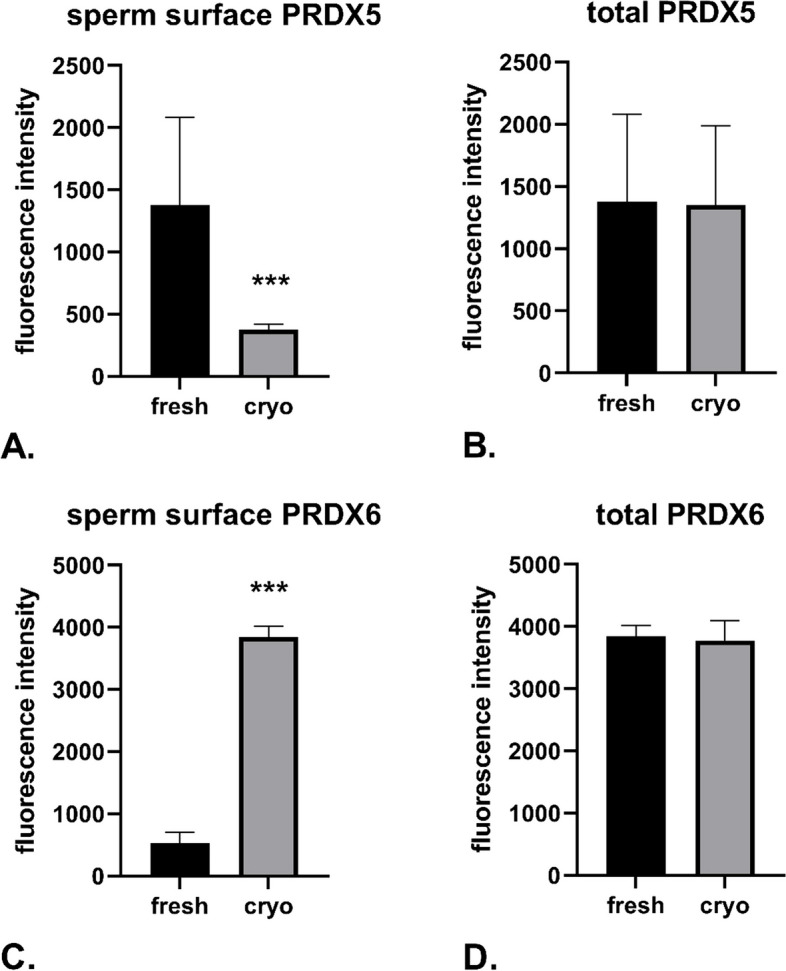
Cryopreservation of bull sperm, crucial for breeding and assisted reproduction, often reduces sperm quality due to oxidative stress. This study examines how oxidative stress during cryopreservation affects peroxiredoxin 5 (PRDX5) and peroxiredoxin 6 (PRDX6) proteins, leading to their translocation and oligomerization in bull sperm. Increased reactive oxygen species (ROS) and nitric oxide (NO) levels were linked to reduced mitochondrial potential, higher DNA fragmentation, and increased membrane fluidity, prompting PRDX5 to move intracellularly and PRDX6 to the cell membrane. Under cryopreservation, these proteins formed high molecular weight oligomers, that may shift from peroxidase to chaperone roles. Their interaction with Toll-like receptor 4 (TLR4) may be key to their intracellular transport. On the other hand, the presence of PRDX5 and PRDX6 in exosomal vesicles suggested a potential mechanism for their transport into sperm cells. Using Imaging Flow Cytometry and various PAGE techniques, the study detected PRDX5 and PRDX6 in different sperm locations and analyzed their oligomer formation. These findings highlight the adaptive roles of PRDX5 and PRDX6 in protecting sperm cells, offering insights that could improve cryopreservation protocols in animal breeding and human reproductive medicine, and advance our understanding of the oxidative stress response in sperm cells.

Keywords: Bull sperm; Cryopreservation; Oxidative stress; PRDX5; PRDX6.


Conflict of interest statement

Declarations. Competing interests: The authors declare no competing interests.

Figures

Fig. 1
Fig. 1
Presence of PRDX5 and PRDX6 on the sperm surface and within the entire sperm cell. Bar charts depict fluorescence intensity of fluorescently labeled anti-PRDX5 antibodies in non-permeabilized (A) and permeabilized (B) sperm samples, as well as anti-PRDX6 antibodies in non-permeabilized (C) and permeabilized (D) sperm samples. Data are presented as the mean ± SD (n = 8 sperm samples). ***p < 0.001
Fig. 2
Fig. 2
Imaging Flow Cytometry images of fresh and cryopreserved sperm, depicting the unstained sperm in Bright Field (A), stained for ROS + presence (B), labeled for PRDX5 on the sperm surface (C), and labeled for PRDX6 on the sperm surface (D). Corresponding Imaging Flow Cytometry plots illustrate sperm populations displaying the presence or absence of PRDX5 and PRDX6 in fresh (E) and cryopreserved (F) samples
Fig. 3
Fig. 3
EV content, size, and PRDX5/PRDX6 expression in SEC fractions from bovine seminal plasma. In bovine seminal plasma SEC fractions 4–7, 72% of the detected particles were membranous (Cell Mask Green positive, CMG +). Of these, 61% expressed at least one EV-related surface marker (CD9 or CD63). The population marked in purple represents CMG + and CD63 + particles, the green population represents CMG + and CD9 + particles, and the yellow population represents CMG + , CD63 + , and CD9 + particles (A). EVs from SEC fractions 4–7 were sized between 100 and 200 nm (B). Western blot analysis of proteins separated by non-reducing SDS-PAGE, using anti-PRDX5 (C) and anti-PRDX6 (D) antibodies, demonstrated the presence of PRDX5 in the exosomal vesicle (EV) fraction, with no detectable signal in the exosome-depleted seminal plasma (SP). Arrows indicate 8 reactive bands for PRDX5 with wide MW range for PRDX5 in the EV fraction. In contrast, PRDX6 was detected in both the EV fraction (EVs) and the exosome-depleted seminal plasma (SP), with arrows indicating 3 reactive bands for PRDX6 with high MW for PRDX6, although the signal was weaker in both
Fig. 4
Fig. 4
Identification of PRDX5 and PRDX6 complexes with TLR4 in sperm cells. Anti-TLR4 monoclonal antibodies were immobilized on a HiTrap protein A HP column (Cytiva) to capture TLR4-PRDX complexes from sperm lysates. The chromatogram highlights the fraction containing protein complexes associated with immobilized TLR4 (A). After elution and concentration, the immunoprecipitated proteins were analyzed by non-reducing SDS-PAGE, followed by Western blotting. Four distinct reactive bands corresponding to PRDX5 were detected above 150 kDa (B), while 12 reactive bands corresponding to PRDX6 were observed, ranging from 17 kDa to above 250 kDa (C). The reactive bands are indicated by red arrows. These results confirm the association of PRDX5 and PRDX6 with TLR4, indicating the formation of TLR4-PRDX complexes in sperm cells
Fig. 5
Fig. 5
Graphs with corresponding representative Western blot analyses of PRDX5 under various electrophoretic conditions: (A) Native PAGE, showing a significant increase in the normalized intensity of the native band after cryopreservation (p < 0.01); (B) Non-denaturing PAGE, showing an increase in the normalized intensity of the native band after cryopreservation (p < 0.001). Lines f1-f4 correspond to fresh sperm samples, while c1-c4 correspond to cryopreserved sperm samples
Fig. 6
Fig. 6
Graphs with corresponding representative Western blot analyses of PRDX5 under various electrophoretic conditions: Standard SDS-PAGE, with no changes in 17.5 kDa band intensity post-cryopreservation (A); Non-reducing SDS-PAGE, showing significant increases in the intensities of the > 250 kDa (B), 100 kDa (C) and 10 kDa (D) bands after cryopreservation (p < 0.05). Lines f1-f4 correspond to fresh sperm samples, while c1-c4 correspond to cryopreserved sperm samples
Fig. 7
Fig. 7
Graphs with corresponding representative Western blot analyses of PRDX6 under various electrophoretic conditions: (A) Native PAGE, showing no changes in the intensity of bands after cryopreservation; (B) Non-denaturing PAGE, showing no changes in the intensity of band post-cryopreservation; Lines f1-f4 correspond to fresh sperm samples, while c1-c4 correspond to cryopreserved sperm samples
Fig. 8
Fig. 8
Graphs with corresponding representative Western blot analyses of PRDX6 under various electrophoretic conditions: (A) Standard SDS-PAGE, with no changes in the intensity of the single 25 kDa band after cryopreservation; Non-reducing SDS-PAGE, showing a significant increase in the intensity of > 250 kDa (B) and 50 kDa (C) bands after cryopreservation (p < 0.05). Lines f1-f4 correspond to fresh sperm samples, while c1-c4 correspond to cryopreserved sperm samples, while c1-c4 correspond to cryopreserved sperm samples

References

    1. Mostek A, Dietrich MA, Słowińska M, Ciereszko A. Cryopreservation of bull semen is associated with carbonylation of sperm proteins. Theriogenology. 2017;92:95–102. Available from: http://linkinghub.elsevier.com/retrieve/pii/S0093691X17300110 - PubMed
    1. Xin L, Guo Y, Zhao HB, Yu HM, Hou L. Peroxiredoxin 6 translocates to the plasma membrane of human sperm under oxidative stress during cryopreservation. Cryobiology. 2021;100:158–63. - PubMed
    1. Rezaei N, Mohammadi M, Mohammadi H, Khalatbari A, Zare Z. Acrosome and chromatin integrity, oxidative stress, and expression of apoptosis-related genes in cryopreserved mouse epididymal spermatozoa treated with L-Carnitine. Cryobiology. 2020;95:171–6. - PubMed
    1. Aitken RJ, Gibb Z, Baker MA, Drevet J, Gharagozloo P. Causes and consequences of oxidative stress in Spermatozoa. Reprod Fertil Dev. 2016;28:1. - PubMed
    1. Knoops B, Goemaere J, Van Der Eecken V, Declercq JP. Peroxiredoxin 5: Structure, mechanism, and function of the mammalian atypical 2-cys peroxiredoxin. Antioxid Redox Signal. 2011;15:817–29. - PubMed

LinkOut - more resources