. 2025 Jan 9;44(1):10.
doi: 10.1186/s13046-024-03267-6.

ONC213: a novel strategy to resensitize resistant AML cells to venetoclax through induction of mitochondrial stress

Affiliations

ONC213: a novel strategy to resensitize resistant AML cells to venetoclax through induction of mitochondrial stress

Jenna L Carter et al. J Exp Clin Cancer Res. .

Abstract

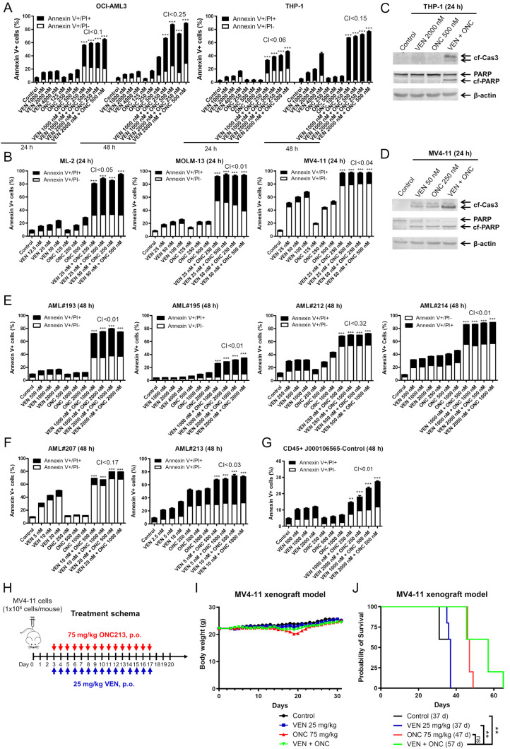
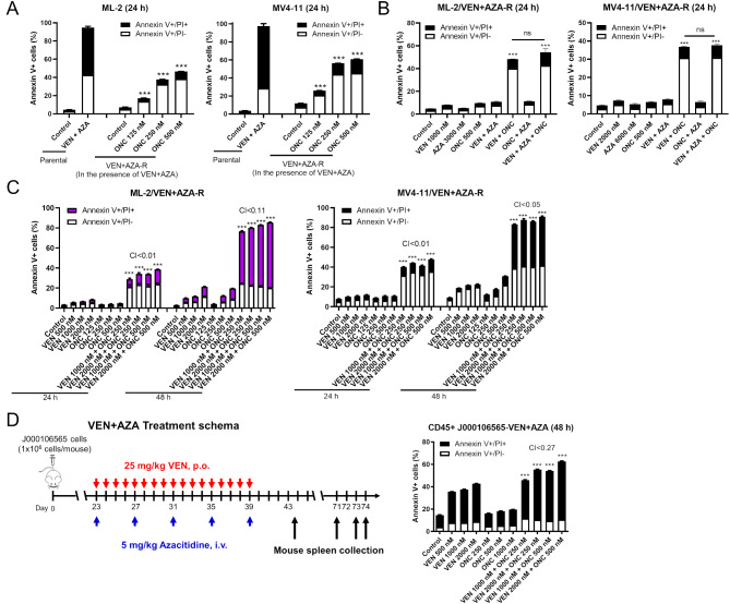
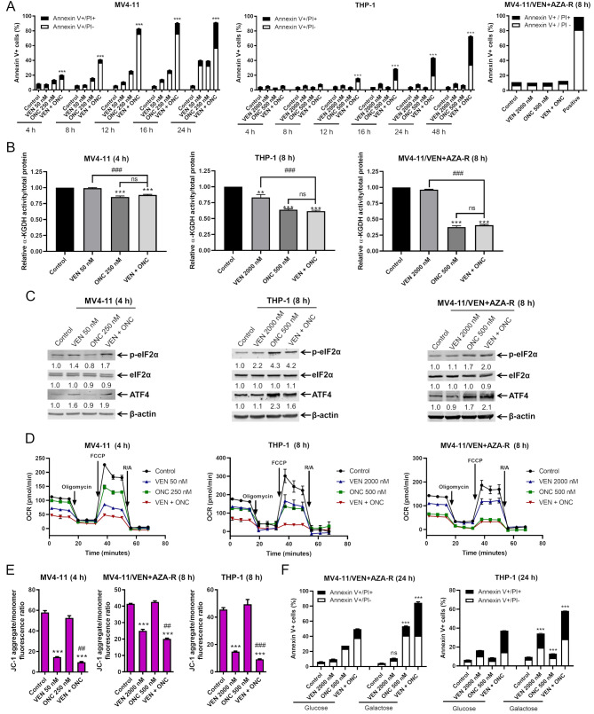
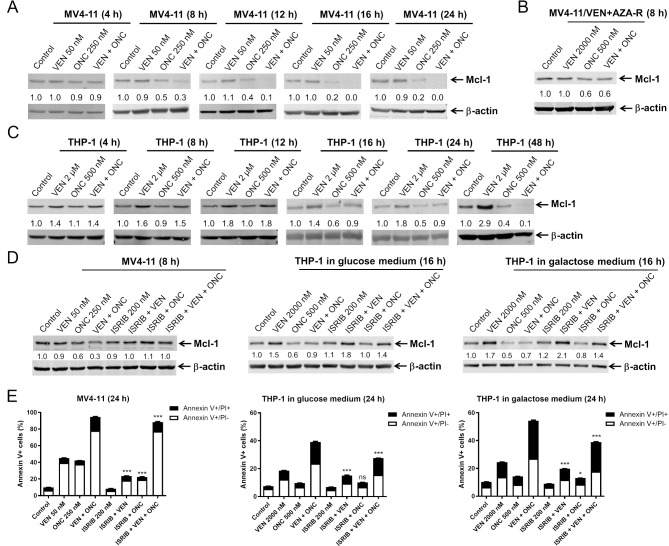
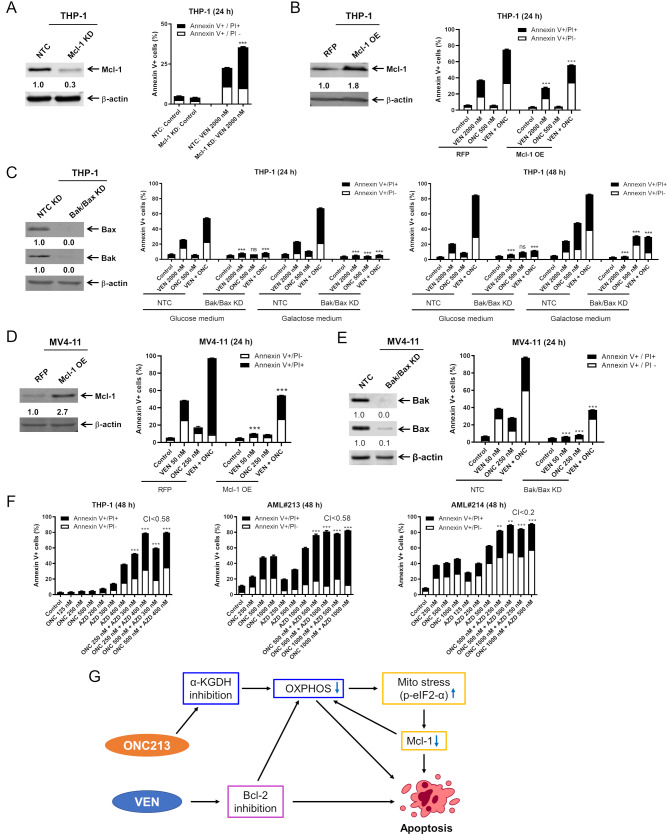
Background: Venetoclax + azacitidine is a frontline treatment for older adult acute myeloid leukemia (AML) patients and a salvage therapy for relapsed/refractory patients who have been treated with intensive chemotherapy. While this is an important treatment option, many patients fail to achieve complete remission and of those that do, majority relapse. Leukemia stem cells (LSCs) are believed to be responsible for AML relapse and can be targeted through oxidative phosphorylation reduction. We previously reported that ONC213 disrupts oxidative phosphorylation and decreases Mcl-1 protein, which play a key role in venetoclax resistance. Here we investigated the antileukemic activity and underlying molecular mechanism of the combination of ONC213 + venetoclax against AML cells.

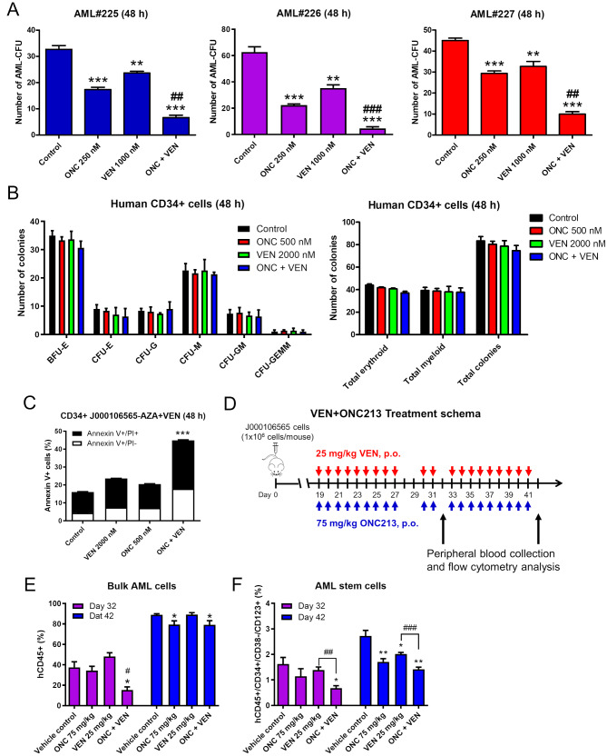
Methods: Flow cytometry was used to determine drug-induced apoptosis. Protein level changes were determined by western blot. An AML cell line-derived xenograft mouse model was used to determine the effects of ONC213 + venetoclax on survival. A patient-derived xenograft (PDX) mouse model was used to determine drug effects on CD45+/CD34+/CD38-/CD123 + cells. Colony formation assays were used to assess drug effects on AML progenitor cells. Mcl-1 and Bax/Bak knockdown and Mcl-1 overexpression were used to confirm their role in the mechanism of action. The effect of ONC213 + venetoclax on mitochondrial respiration was determined using a Seahorse bioanalyzer.

Results: ONC213 + venetoclax synergistically kills AML cells, including those resistant to venetoclax alone as well as venetoclax + azacitidine. The combination significantly reduced colony formation capacity of primary AML progenitors compared to the control and either treatment alone. Further, the combination prolonged survival in an AML cell line-derived xenograft model and significantly decreased LSCs in an AML PDX model.

Conclusions: ONC213 can resensitize VEN + AZA-resistant AML cells to venetoclax therapy and target LSCs ex vivo and in vivo.

Keywords: Acute myeloid leukemia; Azacitidine; ONC213; Venetoclax.


Conflict of interest statement

Declarations. Ethics approval and consent to participate: Diagnostic blast samples were obtained from the First Hospital of Jilin University. Written informed consent was provided, according to the Declaration of Helsinki. This study was approved by the Human Ethics Committee of The First Hospital of Jilin University (Ethical code # 2019 − 128). All animal experiments involving were approved by the Institutional Animal Care and Use Committee at Wayne State University. Consent for publication: Not applicable. Competing interests: ONC213 was provided by Chimerix, Inc., Durham, NC, 27713, and JEA and VVP are employees of Chimerix, Inc. The rest of the authors declare no competing financial interests.

Figures

Fig. 1
Fig. 1
ONC213 + venetoclax has efficacy against AML cells in vitro and in vivo. (A, B, E, F) AML cells were treated with ONC213 (ONC) and venetoclax (VEN) for 24–48 h. The cells were then stained with annexin V/propidium iodide (PI) and analyzed via flow cytometry. ***indicates p < 0.001 compared to control and single drug treatments. Combination Index (CI) values were calculated using CompuSyn software. CI < 1.0, CI = 1.0, and CI > 1.0 indicate synergistic, additive, and antagonistic effects, respectively. (C&D) Representative western blots of whole cell lysates probed with the indicated antibodies are shown. cf-Cas3, cleaved form caspase 3; cf-PARP, cleaved form PARP. (G) J000106565 PDX cells freshly collected from NSGS mouse spleen were treated with ONC and VEN for 48 h and then stained with anti-human CD45 antibody, annexin V, and PI and analyzed by flow cytometry. Annexin V positive human CD45 + cells are graphed. ** p < 0.01 and *** p < 0.001 compared to control and single drug treatments. (H-J) MV4-11 cells were injected into NSGS mice via tail vein. On day 3, the mice were randomized into treatment arms (n = 5) and treated as indicated in panel H. Mouse body weights are graphed in panel I. The overall survival probability was estimated using the Kaplan-Meier method (panel J). ** p < 0.01
Fig. 2
Fig. 2
ONC213 resensitizes venetoclax-resistant cells to venetoclax. (A) ML-2/VEN + AZA-R and MV4-11/VEN + AZA-R cells cultured in the presence of venetoclax + azacitidine (VEN + AZA) were treated with ONC213 (ONC) for 24 h. The parental cells were also treated with VEN + AZA at concentrations that the resistant cells were cultured in for 24 h. The treated cells were stained with annexin V/PI and analyzed via flow cytometry. *** p < 0.001 compared to control. (B) ML-2/VEN + AZA-R and MV4-11/VEN + AZA-R cells cultured without VEN + AZA for 3 days were treated with VEN, AZA, ONC, or in combination for 24 h. The treated cells were stained with annexin V/PI and analyzed via flow cytometry. *** p < 0.001 compared to control and single drug treatments. ns indicates not significant, comparing VEN + ONC and VEN + AZA + ONC. (C) ML-2/VEN + AZA-R and MV4-11/VEN + AZA-R cells (without VEN + AZA present in the media for 72 h) were treated with VEN, ONC, or combination for 24 and 48 h, and then stained with annexin V/PI and analyzed via flow cytometry. Combination Index (CI) values were calculated using CompuSyn software to determine synergy. CI < 1.0, CI = 1.0, and CI > 1.0 indicate synergistic, additive, and antagonistic effects, respectively. *** p < 0.001 compared to vehicle control and single drug treatments. (D) J000106565 cells were injected intravenously through the tail vein of immunocompromised NSGS mice to generate a patient-derived xenograft model. Human cell engraftment was verified in three randomly selected mice 22 days later by flow cytometry for human CD45+ (hCD45+) cells in their peripheral blood (average 20.23%). On day 23, the mice were randomized (n = 5 mice/group) and treated as shown in the upper panel. Mice were sacrificed based on severity of disease-induced symptoms. Cells were isolated from spleens (VEN + AZA treated mice), treated and then stained with anti-hCD45 antibody, annexin V, and PI and analyzed via flow cytometry. Annexin V positive cells are graphed. ** p < 0.01 and *** p < 0.001 compared to control and single drug treatments. CI values were calculated as described in the description for panel C
Fig. 3
Fig. 3
ONC213 and venetoclax target AML progenitor and stem cells. (A) Primary AML patient samples were cultured with ONC213 (ONC) and venetoclax (VEN), alone or in combination, for 48 h and then plated in methylcellulose and incubated for 2 weeks. The number of surviving AML cells capable of generating leukemia colonies (AML-CFUs) were enumerated. Technical triplicates were performed. Data are presented as mean ± SEM. ** p < 0.01 and *** p < 0.001 compared to vehicle control; ## p < 0.01 and ### p < 0.001 compared to single drug treatments. (B) Human CD34 + cord blood cells were treated with ONC213 and VEN, alone or in combination, for 24 h and then plated in methylcellulose and incubated for 2 weeks. The number of surviving normal hematopoietic cells capable of generating colonies were counted and colonies are presented as mean ± SEM. The number of BFU-E, CFU-E, CFU-G, CFU-M, CFU-GM, and CFU-GEMM is presented as mean ± SEM (left panel). Total erythroid and myeloid colonies are presented as mean ± SEM (right panel). (C) AML cells were isolated from spleens of NSGS mice injected with J000106565 patient-derived AML cells and treated with VEN + AZA. CD34 MicroBeads were used to enrich human CD34 + cells. These cells were treated with VEN, ONC, or the combination for 48 h, and then stained with anti-human CD45 and annexin V/PI and analyzed via flow cytometry. *** p < 0.001 compared to control and single drug treatments. (D-F) NSGS mice were injected with J000106565 patient-derived AML cells. Engraftment was confirmed on day 18. On day 19, mice were randomized into treatment groups (n = 5) and treated as shown in panel D. On days 32 (part way through treatment) and 42 (at the end of treatment) peripheral blood was collected, and cells were stained with anti-human CD45, -CD34, -CD38, and -CD123 antibodies and analyzed via flow cytometry. Bulk AML cells were determined by gating for CD45 + cells (panel E). AML stem cells were determined by gating for CD45dim+/CD34+/CD38-/CD123 + cells (panel F). * p < 0.05 and ** p < 0.01 compared to control. # p < 0.05 compared to single drug treatments (left panel). ## p < 0.01 and ### p < 0.001 compared to VEN treatment (right panel)
Fig. 4
Fig. 4
ONC213 induces a mitochondrial stress response and its combination with venetoclax diminishes mitochondria function in AML cells. (A) MV4-11, THP-1, and MV4-11/VEN + AZA-R cells were treated with venetoclax (VEN), ONC213 (ONC), or ONC + VEN for up to 48 h and then stained with annexin V/PI and analyzed via flow cytometry. *** indicates p < 0.001 compared to control and single drug treatments. (B) MV4-11, THP-1, MV4-11/VEN + AZA-R cells were treated with VEN and ONC, alone or in combination, for 4–8 h. α-ketoglutarate dehydrogenase (α-KGDH) activity was measured. The ratio of α-KGDH activity to total protein was determined and normalized to the vehicle control. ** p < 0.01 and *** p < 0.001 compared to control. ### p < 0.001 compared to VEN. ns not significant compared to ONC. (C) MV4-11, THP-1, and MV4-11/VEN + AZA-R cells were treated with VEN and ONC, alone or in combination, for 4–8 h. Representative western blots of whole cell lysates probed with the indicated antibodies are shown. The fold changes for the densitometry measurements, normalized to β-actin and then compared to vehicle control, are indicated below the corresponding blots. (D) MV4-11, THP-1, and MV4-11/VEN + AZA-R cells were treated with VEN and ONC, alone or in combination, for 4–8 h and then subjected to the cellular mito stress test. OCR was measured following injection with oligomycin A (complex V inhibitor), FCCP (mitochondrial membrane uncoupler), and rotenone + antimycin A (complex I and III inhibitors). (E) MV4-11, THP-1, and MV4-11/VEN + AZA-R cells were treated with VEN and ONC, alone or in combination, for 4–8 h and then stained with JC-1 and analyzed via flow cytometry for aggregate vs. monomer fluorescence ratio. *** p < 0.001 compared to control. ## p < 0.01 and ### p < 0.001 compared to ONC alone and VEN alone. (F) MV4-11/VEN + AZA-R and THP-1 cells were cultured in either glucose or galactose containing media for 48 h and then treated with VEN, ONC, or the combination of ONC + VEN for 24 h. Then the cells were stained with annexin V/PI and analyzed via flow cytometry for apoptosis. ns not significant and *** p < 0.001 compared to the same treatment in glucose media
Fig. 5
Fig. 5
ONC213 + venetoclax induced cell death is partially dependent on Mcl-1. (A-C) MV4-11, THP-1, and MV4-11/VEN + AZA-R cells were treated with VEN and ONC, alone or in combination, for up to 48 h. Representative western blots of whole cell lysates probed with the indicated antibodies are shown. The fold changes for the densitometry measurements, normalized to β-actin and then compared to vehicle control, are indicated below the corresponding blots. (D) MV4-11 cells and THP-1 cells (cultured in glucose or galactose containing media) were treated with VEN, ONC, or ISRIB, alone or in combination, for 8–16 h. Representative western blots of whole cell lysates probed with the indicated antibodies are shown. The fold changes for the densitometry measurements, normalized to β-actin and then compared to vehicle control, are indicated below the corresponding blots. (E) MV4-11 cells and THP-1 cells (cultured in glucose or galactose containing media) were treated with VEN, ONC, or ISRIB, alone or in combination, for 24 h and then stained with annexin V/PI and analyzed via flow cytometry. * p < 0.05, *** p < 0.001, and ns not significant compared to the same drug treatment but without ISRIB
Fig. 6
Fig. 6
Induction of mitochondrial stress via ONC213 supports intrinsic apoptosis activation via venetoclax. (A) CRISPR knockdown (KD) of Mcl-1 and non-target control (NTC) was generated using THP-1 cells. Representative western blots of whole cell lysates probed with the indicated antibodies are shown in the left panel. The fold changes for the Mcl-1 densitometry measurements, normalized to β-actin and then compared to NTC, are indicated. Cells were treated with vehicle control or venetoclax (VEN) for 24 h. Flow cytometry results of annexin V/PI staining are show in the right panel. *** p < 0.001 compared to NTC treated with VEN. (B) THP-1 RFP (red fluorescent protein) and Mcl-1 OE (overexpression) whole cell lysates were subjected to western blot analysis. The fold changes for the Mcl-1 densitometry measurements, normalized to β-actin and then compared to RFP, are indicated. Cells were treated with vehicle control, VEN, ONC, or in combination for 24 h. Flow cytometry results of annexin V/PI staining are show in the right panel. *** p < 0.001 compared to RFP cells under the same treatment conditions. (C) THP-1 NTC and Bak/Bax KD whole cell lysates were subjected to western blot analysis. The fold changes for the Bax and Bak densitometry measurements, normalized to β-actin and then compared to NTC, are indicated. THP-1 NTC and Bak/Bax KD cells were cultured in either glucose or galactose containing media for 48 h and then treated with VEN, ONC213, or the combination of ONC + VEN for 24–48 h. Then the cells were stained with annexin V/PI and analyzed via flow cytometry. *** p < 0.001 and ns not significant compared to NTC cells under the same treatment conditions. (D) MV4-11 RFP and Mcl-1 OE whole cell lysates were subjected to western blot analysis. The fold changes for the Mcl-1 densitometry measurements, normalized to β-actin and then compared to RFP, are indicated. Cells were treated with vehicle control, VEN, ONC, or in combination for 24 h. Flow cytometry results of annexin V/PI staining are show in the right panel. *** p < 0.001 compared to RFP cells under the same treatment conditions. (E) MV4-11 NTC and Bak/Bax KD whole cell lysates were subjected to western blot analysis. The fold changes for the Bax and Bak densitometry measurements, normalized to β-actin and then compared to NTC, are indicated. MV4-11 NTC and Bak/Bax KD cells were cultured in glucose containing media for 48 h and then treated with VEN, ONC, or the combination of ONC + VEN for 24 h. Then the cells were stained with annexin V/PI and analyzed via flow cytometry. *** p < 0.001 compared to NTC cells under the same treatment conditions. (F) THP-1 cells and primary AML patient samples AML#213 and AML#214 were treated with VEN, ONC, or VEN + ONC for 48 h. The cells were then stained with annexin V/PI and subjected to flow cytometry analyses. Combination Index (CI) values were calculated using CompuSyn software to determine synergy. CI < 1.0, CI = 1.0, and CI > 1.0 indicate synergistic, additive, and antagonistic effects, respectively. ** p < 0.01 and *** p < 0.001 compared to control and single drug treatments. (G) Proposed mechanism of action of ONC + VEN. ONC213 targets oxidative phosphorylation via inhibition of α-KGDH which subsequently induces mitochondrial stress pathways (p-eIF2-α) and reduces levels of Mcl-1. Mcl-1 is a known contributor to resistance to venetoclax and reduction of Mcl-1 activates the intrinsic apoptosis pathway and contributes to reduction of oxidative phosphorylation, enhancing the antileukemic activity of venetoclax against AML

Similar articles

Cited by

References

    1. SEER Cancer Stat Facts. Acute Myeloid Leukemia Bethesda, MD: National Cancer Institute; [Available from: https://seer.cancer.gov/statfacts/html/amyl.html]
    1. Pulte D, Jansen L, Brenner H. Changes in long term survival after diagnosis with common hematologic malignancies in the early 21st century. Blood cancer J. 2020;10(5):56. - PMC - PubMed
    1. Krug U, Büchner T, Berdel WE, Müller-Tidow C. The treatment of elderly patients with acute myeloid leukemia. Deutsches Arzteblatt Int. 2011;108(51–52):863–70. - PMC - PubMed
    1. Bradbury DA, Zhu YM, Russell NH. Bcl-2 expression in acute myeloblastic leukaemia: relationship with autonomous growth and CD34 antigen expression. Leuk Lymphoma. 1997;24(3–4):221–8. - PubMed
    1. Delia D, Aiello A, Soligo D, Fontanella E, Melani C, Pezzella F, et al. bcl-2 proto-oncogene expression in normal and neoplastic human myeloid cells. Blood. 1992;79(5):1291–8. - PubMed

MeSH terms