. 2025 Jan;286(1):e70026.
doi: 10.1002/jmor.70026.

Vascular and Osteological Morphology of Expanded Digit Tips Suggests Specialization in the Wandering Salamander (Aneides vagrans)

Affiliations

Vascular and Osteological Morphology of Expanded Digit Tips Suggests Specialization in the Wandering Salamander (Aneides vagrans)

Christian E Brown et al. J Morphol. 2025 Jan.

Abstract

For over a century researchers have marveled at the square-shaped toe tips of several species of climbing salamanders (genus Aneides), speculating about the function of large blood sinuses therein. Wandering salamanders (Aneides vagrans) have been reported to exhibit exquisite locomotor control while climbing, jumping, and gliding high (88 m) within the redwood canopy; however, a detailed investigation of their digital vascular system has yet to be conducted. Here, we describe the vascular and osteological structure of, and blood circulation through, the distal regions of the toes of A. vagrans using histology in tandem with live-animal videos. Specifically, we sectioned a toe of A. vagrans at 0.90 μm, embedded it in Spurrs resin, and stained the tissue with toluidine blue. An additional three toes were sectioned at 10 μm, embedded in paraffin, and after sectioning and mounting, treated with Verhoeff and Quad stains. For living salamanders, we recorded real-time videos of blood flowing within individual toes upon a translucent surface oriented both horizontally (0°) and vertically (90°) to simulate both prostrate and vertical clinging scenarios, then analyzed the image sequences using ImageJ. We found that the vascularized toe tips have one large sinus cavity that is divided more proximally into two chambers via a septum, and there are mucous and granular glands in the dorsal and dorsolateral integument of the digit tips. Live-animal trials revealed variable sinus-filling both within and between toes, seemingly associated with variable pressure applied to the substrate when standing, stepping, clinging, and climbing. We conclude that A. vagrans, and likely other climbing salamanders, can functionally fill, trap, and drain the blood in their vascularized toe tips to optimize attachment, detachment, and complex arboreal locomotion (e.g., landing after gliding flight). Such an adaptation could provide insights for bioinspired designs.

Keywords: arboreal; blood sinus; locomotion; mucous gland; toe.


Conflict of interest statement

The authors declare no conflicts of interest.

Figures
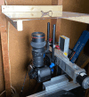
Figure 1
Figure 1
Custom built salamander‐viewing platform and camera slide used to image live Aneides vagrans. The viewing platform can be positioned vertically or horizontally (as shown here) and secured during trials. The viewing platform was made of clear acrylic such that images from the ventral view can be easily captured, the latter being the most expansive single view for visualizing the entire digital vascular system.
Figure 2
Figure 2
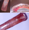
Still frame images of Digit III, the middle toe, of a live Aneides vagrans. Images were taken with the salamander in a neutral posture on a flat surface from (A) dorsal, (B) lateral, and (C) ventral views. These images show the digital sinus and the bifurcated terminal phalanx of a living salamander; the structure of these features was corroborated by our histological preparations, shown in Figures 3 and 4.
Figure 3
Figure 3
Detailed schematic representation of the toe tip of Aneides vagrans. Schematic representations are shown in ventral and lateral views. Schematics show the epidermis (blue line), terminal phalanx (light pink), joint (magenta), connective tissue (beige), blood sinus (brown), artery (red), blood vessels (orange‐brown), granular glands (purple ovals), and mucus glands (pink ovals). Lines drawn through the toe at four distinct points along the toe tip (A–D) indicate the points at which the cross‐sections featured in Figure 4 were taken.
Figure 4
Figure 4
Histology of Aneides vagrans toe. Photomicrographs of 10 μm (A, B, C, and D) cross sections of Digit III of the hind foot. Photomicrograph panels A and D represent slides stained with Quad stain, while photomicrographs C and D are stained with Verhoeff stain. Series A through D represent slides moving from most distal (A) to most proximal (D) along the distal phalanx before the most distal interphalangeal joint. Anatomical components visible: BS, blood sinus; CTS, connective tissue septum; DER, dermis; DP, distal phalanx; E, tissue staining positive for elastin; EPI, epidermis; GG, granular glands; MC, medullary cavity; MG, mucous glands; VE, ventral extension of the distal phalanx. Scale bars are 100 μm.
Figure 5
Figure 5
Bar graph comparing the average change in redness (blue bars) from images of the digital sinuses of A. vagrans under various locomotory conditions. On average, the digital redness is highest in the last frame before toe off and lowest at 300 frames, or 10 s, before toe off. Standard deviations are shown as error bars (black).
Figure 6
Figure 6
Still frame images of Digit III, the middle‐most toe, of a live Aneides vagrans when (A) drained of (top) vs. (B) engorged (bottom) with blood. Both (A and B) show the same toe of the same salamander during the same experimental trial, the images being separated by ~25 s. The artery supplying the digital sinus increased in visible width by ~23 μm, or ~25%, and the toe tip extended proximally on the ventral side, with distance measurements at the longest section of the toe tip increasing proximally by ~45 μm, or ~5%. See Movie S2.

Similar articles

References

    1. Adams, D. C. , Korneisel D., Young M., and Nistri A.. 2017. “Natural History Constrains the Macroevolution of Foot Morphology in European Plethodontid Salamanders.” American Naturalist 190, no. 2: 292–297. 10.1086/692471. - DOI - PubMed
    1. Adams, D. C. , and Nistri A.. 2010. “Ontogenetic Convergence and Evolution of Foot Morphology in European Cave Salamanders (Family: Plethodontidae).” BMC Evolutionary Biology 10: 216. 10.1186/1471-2148-10-216. - DOI - PMC - PubMed
    1. Afferante, L. , Heepe L., Casdorff K., Gorb S., and Carbone G.. 2016. “A Theoretical Characterization of Curvature Controlled Adhesive Properties of Bio‐Inspired Membranes.” Biomimetics 1, no. 1: 3. 10.3390/biomimetics1010003. - DOI
    1. Aretz, J. , Brown C. E., and Deban S. M.. 2021. “Vertical Locomotion in the Arboreal Salamander Aneides Vagrans .” Zoology 316: 72–79. 10.1111/jzo.12934. - DOI
    1. Baken, E. K. , and Adams D. C.. 2019. “Macroevolution of Arboreality in Salamanders.” Ecology and Evolution 9, no. 12: 7005–7016. 10.1002/ece3.5267. - DOI - PMC - PubMed

LinkOut - more resources