. 2025 Dec;47(1):2449195.
doi: 10.1080/0886022X.2024.2449195. Epub 2025 Jan 8.

Identification of biomarkers for chronic renal fibrosis and their relationship with immune infiltration and cell death

Affiliations

Identification of biomarkers for chronic renal fibrosis and their relationship with immune infiltration and cell death

Jiaqi Shi et al. Ren Fail. 2025 Dec.

Abstract

Background: Chronic kidney disease (CKD) represents a significant global public health challenge. This study aims to identify biomarkers of renal fibrosis and elucidate the relationship between unilateral ureteral obstruction (UUO), immune infiltration, and cell death.

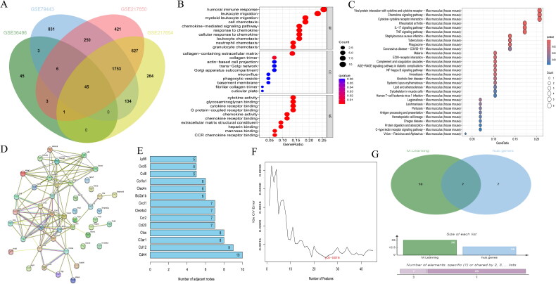
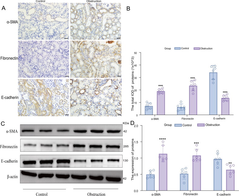
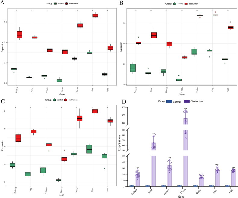
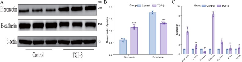
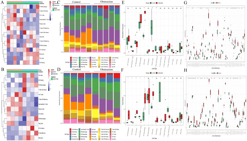
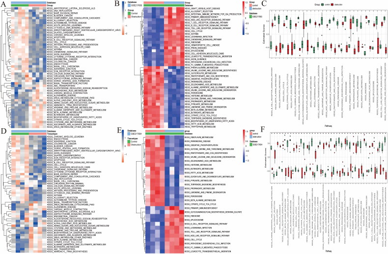
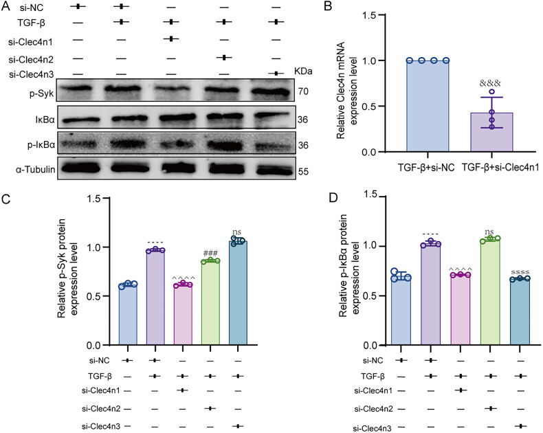
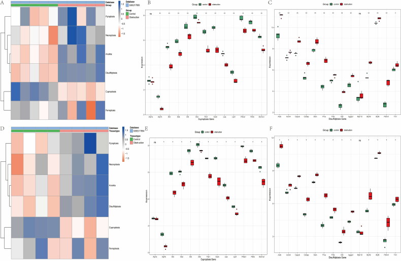
Methods: Gene expression matrices for UUO were retrieved from the gene expression omnibus (GSE36496, GSE79443, GSE217650, and GSE217654). Seven genes identified through Protein-Protein Interaction (PPI) network and Support Vector Machine-Recursive Feature Elimination (SVM-RFE) analysis were validated using qRT-PCR in both in vivo and in vitro UUO experiments. WB assays were employed to investigate the role of Clec4n within NF-κB signaling pathway in renal fibrosis. The composition of immune cells in UUO was assessed using CIBERSORT, and gene set variant analysis (GSVA) was utilized to evaluate prevalent signaling pathways and cell death indices.

Results: GO and KEGG enrichment analyses revealed numerous inflammation-related pathways significantly enriched in UUO conditions. Bcl2a1b, Clec4n, and Col1a1 were identified as potential diagnostic biomarkers for UUO. Analysis of immune cell infiltration indicated a correlation between UUO and enhanced mast cell activation. Silencing Clec4n expression appeared to mitigate the inflammatory response in renal fibrosis. GSVA results indicated elevated inflammatory pathway scores in UUO, with significant differences in disulfiram and cuproptosis scores compared to those in the normal murine kidney group.

Conclusion: Bcl2a1b, Clec4n, and Col1a1 may serve as biomarkers for diagnosing UUO. UUO development is closely linked to immune cell infiltration, activation of inflammatory pathways, disulfiram, and cuproptosis processes.

Keywords: Chronic kidney fibrosis; bioinformatics; biomarkers; cell death; immune infiltration.


Conflict of interest statement

No potential conflict of interest was reported by the author(s).

Figures

Figure 1.
Figure 1.
DEGs based on PPI network, machine learning analysis and functional enrichment analysis in UUO. (A) Wayne diagram showing DEGs shared by four datasets, GSE36496, GSE79443, GSE217650, and GSE217654; (B) Gene ontology (GO) enrichment analysis; (C) KEGG enrichment analysis; (D) PPI network based on the PPI network of DEGs; (E) Top 14 genes with the highest connectivity in the PPI network; (F) Graph of biomarkers screened by the SVM-RFE algorithm; (G) Wayne’s plot showing seven diagnostic biomarkers shared by PPI and SVM-RFE.
Figure 2.
Figure 2.
Experimental validation of UUO model by immunohistochemistry and Western blot. (A) Immunohistochemistry of α-SMA, Fibronectin and E-cadherin fibrosis indexes in the control group (n = 6) and the obstruction group (n = 6) (magnification: ×400, scale bar = 100 µm); (B)Analysis of IOD values of fibrosis indexes in both groups; (C) Analysis of protein blotting of α-SMA, Fibronectin and E-cadherin in control and obstruction group; β-actin was used as a loading control; The data are presented as the mean ± standard deviation of three independent experiments; **P < 0.01, ***P < 0.001, ****P < 0.0001 vs. Control.
Figure 3.
Figure 3.
Real-time quantitative PCR validation of diagnostic biomarkers for UUO. (A) Expression of DEGs in the GSE79443 dataset; (B) Expression of DEGs in the GSE217650 dataset; (C) Expression of DEGs in the GSE217654 dataset; (D) The expression levels of seven DEGs in kidney tissues of the control group (n = 6) and the obstruction group (n = 6) were measured using qRT-PCR, with GAPDH serving as the housekeeping gene control; The data are presented as the mean ± standard deviation of three independent experiments; **** p < 0.0001 vs. Control.
Figure 4.
Figure 4.
Validation of 7 differential genes in fibrotic mouse renal tubular epithelial cells. (A and B) Analysis of protein blotting of Fibronectin and E-cadherin in control and TGF-β group; (C) The expression levels of seven DEGs in the mouse renal fibrosis cell model were measured using qRT-PCR, with GAPDH serving as the housekeeping gene control; The data are presented as the mean ± standard deviation of three independent experiments; not significant: ns; ***p < 0.001, **p < 0.01 vs. Control.
Figure 5.
Figure 5.
Relationship between UUO and immune cell infiltration and immune checkpoint molecules. (A and B) The heatmaps of the distribution of immune cells in the control and obstruction groups in the two datasets, GSE217650 and GSE217654; (C and D) The characterization of immune cells by the CIBERSORT method in the two datasets; (E and F) The comparison of immune cells in the control and obstruction groups in the two datasets; (G and H) The comparison of the surface markers of immune cells in the control and obstruction groups in the two datasets.
Figure 6.
Figure 6.
GSVA Signaling pathway variant set analysis. (A and D) The common heatmap of the relevant signaling pathways in the GSE217650 dataset and GSE217654 dataset; (B and E) The heatmap after difference analysis of the relevant signaling pathways in the GSE217650 dataset and GSE217654 dataset; (C and F) Comparison of enrichment scores of relevant signaling pathways between control and obstruction groups under GSVA analysis in the GSE217650 dataset and GSE217654 dataset.
Figure 7.
Figure 7.
WB And qRT-PCR experiments verified the impact of Clec4n silencing on NF-κB signaling in renal fibrosis. (A, C, and D) The protein expression level of p-Syk and p-IκBα were measured by Western blot. α-Tubulin was taken as the loading control. (B) The efficiency of Clec4n knockdown was determined by qRT-PCR. GAPDH was used as a housekeeping gene control. The data are presented as the mean ± standard deviation of three independent experiments. ****P < 0.0001 vs. si-NC; ^^^^P < 0.0001 vs. si-NC + TGF-β; ###P < 0.001 vs. si-NC + TGF-β; $$$$P < 0.0001 vs. si-NC+ TGF-β; &&&P < 0.001 vs. TGF-β + si-NC, and not significant (ns).
Figure 8.
Figure 8.
Analysis of cell death-related signaling pathways by GSVA. (A and D) The heatmap of the enrichment scores related to pyroptosis, necroptosis, anoikis, disulfidptosis, cuproptosis and ferroptosis in the GSE217650 and GSE217654 datasets; (B and E) Comparison of the enrichment scores of cuproptosis-related signaling pathways between control and obstruction groups in the GSE217650 and GSE217654 datasets; (C and F) Comparison of the enrichment scores of disulfidptosis-related signaling pathways between control and obstruction groups in the GSE217650 and GSE217654 datasets.

Similar articles

Cited by

References

    1. Rongshuang H, Ping F, Liang M.. Kidney fibrosis: from mechanisms to therapeutic medicines. Signal Transduct Target Ther; 2023;8(1):129. - PMC - PubMed
    1. Miao H, Wang Y-N, Su W, et al. . Sirtuin 6 protects against podocyte injury by blocking the renin-angiotensin system by inhibiting the Wnt1/β-catenin pathway. Acta Pharmacol Sin. 2023;45(1):137–149. - PMC - PubMed
    1. Yuan Q, Tang B, Zhang C.. Signaling pathways of chronic kidney diseases, implications for therapeutics. Signal Transduct Target Ther. 2022;7(1):182. doi: 10.1038/s41392-022-01036-5. - DOI - PMC - PubMed
    1. Huang J, Liu Y, Shi M, et al. . Empagliflozin attenuating renal interstitial fibrosis in diabetic kidney disease by inhibiting lymphangiogenesis and lymphatic endothelial-to-mesenchymal transition via the VEGF-C/VEGFR3 pathway. Biomed Pharmacother. 2024;180:117589. - PubMed
    1. Peng Y, Li L, Shang J, et al. . Macrophage promotes fibroblast activation and kidney fibrosis by assembling a vitronectin-enriched microenvironment. Theranostics. 2023;13(11):3897–3913. doi: 10.7150/thno.85250. - DOI - PMC - PubMed

MeSH terms