. 2025 Dec;47(1):2447801.
doi: 10.1080/0886022X.2024.2447801. Epub 2025 Jan 8.

Piezo1 aggravates ischemia/reperfusion-induced acute kidney injury by Ca2+-dependent calpain/HIF-1α/Notch signaling

Affiliations

Piezo1 aggravates ischemia/reperfusion-induced acute kidney injury by Ca2+-dependent calpain/HIF-1α/Notch signaling

Xiaoting Chen et al. Ren Fail. 2025 Dec.

Abstract

Macrophages play a vital role in the inflammation and repair processes of ischemia/reperfusion-induced acute kidney injury (IR-AKI). The mechanosensitive ion channel Piezo1 is significant in these inflammatory processes. However, the exact role of macrophage Piezo1 in IR-AKI is unknown. The main purpose of this study was to determine the role of macrophage Piezo1 in the injury and repair process in IR-AKI. Genetically modified mice with targeted knockout of Piezo1 in myeloid cells were established, and acute kidney injury was induced by bilateral renal vascular clamping surgery. Additionally, hypoxia treatment was performed on bone marrow-derived macrophages in vitro. Our data indicate that Piezo1 is upregulated in renal macrophages in mice with IR-AKI. Myeloid Piezo1 knockout provided protective effects in mice with IR-AKI. Mechanistically, the regulatory effects of Piezo1 on macrophages are at least partially linked to calpain signaling. Piezo1 activates Ca2+-dependent calpain signaling, which critically upregulates HIF-1α signaling. This key pathway subsequently influences the Notch and CCL2/CCR2 pathways, driving the polarization of M1 macrophages. In conclusion, our findings elucidate the biological functions of Piezo1 in renal macrophages, underscoring its role as a crucial mediator of acute kidney injury. Consequently, the genetic or pharmacological inhibition of Piezo1 presents a promising strategy for treating IR-AKI.

Keywords: IR-AKI; Piezo1; calpain; hypoxia; macrophage; polarization.


Conflict of interest statement

No potential conflict of interest was reported by the author(s).

Figures

None
Graphical abstract
Figure 1.
Figure 1.
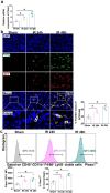
Piezo1 is upregulated in macrophages after IR-AKI. (a) Relative mRNA expression of Piezo1 in normal or injured kidney tissues normalized to that of 18S (n = 6). (b) Double immunofluorescence staining showing F4/80 (green) with RFP (red) in kidney slices from Piezo1td/Tdt mice at the indicated time points (n = 6). Nuclei are stained with DAPI (blue). Scale bar: 50 μm. (c) Representative flow cytometric images illustrating the percentage and Geo-MFI levels of Piezo1+ cells in macrophages (CD45+ CD11b+ F4/80+ Ly6G- live cells) from WT mice subjected to sham or renal IR injury surgery (n = 6). One-way ANOVA with the Bonferroni was conducted in a–c. Error bars represent the mean ± SEM. *P < 0.05. RFP, red fluorescent protein; td/Tdt: tdTomato; WT, wild-type; DAPI, 4’,6’-diamidino-2-phenylindole; GEO-MFI, geometric mean fluorescence intensity; IR, ischemia/reperfusion.
Figure 2.
Figure 2.
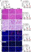
Myeloid-specific Piezo1 deficiency attenuates IR-AKI. Piezo1fl/fl or Piezo1ΔLysM mice were subjected to sham or renal IR injury surgery. (a–b) SCr and BUN concentrations in different groups of mice (n = 6). (c–d) Representative images of HE and PAS staining showing renal morphology and quantitative assessment of renal tubular injury in different groups of mice. (n = 6). Scale bars: 50 μm. (e) Representative images and quantification of immunofluorescence staining of KIM-1 in kidneys from different groups of mice (n = 6). Scale bar: 50 μm. (f) Representative in situ TUNEL assay and quantitative assessment of nephron cell death in kidneys of different groups of mice (n = 6). Nuclei are stained with DAPI (blue). Scale bar: 50 μm. Two-way ANOVA with the Bonferroni was conducted in a–f. Error bars represent the mean ± SEM. *P < 0.05. SCr, serum creatinine; BUN, blood urea nitrogen; HE, hematoxylin and eosin; PAS, periodic acid-Schiff; TUNEL, terminal deoxynucleotidyl transferase dUTP nick-end labeling. DAPI, 4’,6’-diamidino-2-phenylindole; IR, ischemia/reperfusion.
Figure 3.
Figure 3.
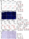
Piezo1 is involved in IR-AKI by regulating macrophages. Piezo1fl/fl or Piezo1ΔLysM mice were subjected to sham or renal IR injury surgery. (a) Representative flow cytometry images showing quantitative data on the expression, GEO-MFI, and percentage of neutrophils (CD45+ CD11b+ Ly6G+ cells) in kidney tissues from different groups of mice (n = 6). (b) Representative images and quantification of immunofluorescence staining of Ly6G (green) in kidneys from different groups of mice (n = 6). Nuclei are stained with DAPI (blue). Scale bar: 50 μm. (c) Representative flow cytometry images showing the expression and percentage of F4/80low+ and F4/80high+ macrophages in kidney tissues from different groups of mice (n = 6). (d) Representative images and quantification of immunohistochemical staining for CD68 in kidneys from different groups of mice (n = 6). Nuclei are stained with DAPI (blue). Scale bar: 50 μm. Two-way ANOVA with the Bonferroni was conducted a–e. Error bars represent the mean ± SEM. *P < 0.05. GEO-MFI, geometry-mean fluorescence intensity; DAPI, 4’,6’-diamidino-2-phenylindole; IR, ischemia/reperfusion.
Figure 4.
Figure 4.
Piezo1 exacerbates IR-AKI by promoting M1 macrophage polarization. Piezo1fl/fl or Piezo1ΔLysM male mice were subjected to sham or renal IR injury surgery. (a–c) Representative flow cytometric images illustrating the percentage and Geo-MFI levels of CD86+, MHC-II+, and CD206+ cells among F4/80low macrophages in kidneys from different groups of mice (n = 6). (d–e) Representative images and quantification of immunofluorescence staining of CD80(M1 marker) and CD163 (M2 marker) in kidneys from different groups of mice (n = 6). Nuclei are stained with DAPI (blue). Scale bar: 50 μm. (f) Relative mRNA expression levels of IL6, TNF-α, IL1β, Arg1, and MRC1 in kidneys from different groups of mice normalized to that of 18S. The heatmap shows the logarithmic values of the gene expression levels (n = 6). Two-way ANOVA with the Bonferroni was conducted in a–f. Error bars represent the mean ± SEM. *P < 0.05. GEO-MFI, geometry-mean fluorescence intensity; DAPI, 4’,6’-diamidino-2-phenylindole; IR, ischemia/reperfusion.
Figure 5.
Figure 5.
Piezo1 promotes M1 macrophage polarization via calpain activity. (a–b) Representative flow cytometric images illustrating the percentage and Geo-MFI levels of CD86+, and MHC-II+ cells in hypoxia-induced BMDMs (F4/80+ live cells) from Piezo1fl/fl and Piezo1ΔLysM mice (n = 4). (c) Calpain activity in kidneys from Piezo1fl/fl and Piezo1ΔLysM mice subjected to sham or renal IR injury surgery (n = 6). (d) Calpain activity in hypoxia-induced BMDMs from Piezo1fl/fl and Piezo1ΔLysM mice (n = 4). (e) Calpain activity in BMDMs treated with 10 μM Yoda1 (n = 4). (f) Calpain activity in hypoxia-induced BMDMs with or without 2 μM PD150606 (n = 4). (g) Calpain activity in stretch-induced BMDMs (n = 4). (h) Calpain activity in stretch-induced BMDMs with or without 2 μM PD150606 (n = 4). (i–j) Representative flow cytometric images illustrating the percentage and Geo-MFI levels of CD86+, and MHC-II+ cells in hypoxia-induced BMDMs (CD11b+ F4/80+ live cells) from WT mice after treatment with Yoda1 or PD150606 (n = 4). Two-way ANOVA with the Bonferroni was conducted in a–e, and g; One-way ANOVA with the Bonferroni was conducted in f, and h–j. Error bars represent the mean ± SEM. *P < 0.05. GEO-MFI, geometry-mean fluorescence intensity; WT, wild-type; DAPI, 4’,6’-diamidino-2-phenylindole.
Figure 6.
Figure 6.
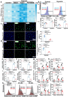
HIF-1α, Notch and CCL2/CCR2 signaling is involved in Piezo1-mediated M1 macrophage polarization. (a) Normalized heatmap of the relative expression levels of mRNAs in BMDMs from WT mice subjected to different treatments normalized to that of 18S (n = 4). (b) Representative flow cytometric images illustrating the percentage and Geo-MFI levels of CCR2+ cells among BMDMs (F4/80+ live cells) from Piezo1fl/fl and Piezo1ΔLysM mice (n = 4). (c–d) Representative images and quantification of immunofluorescence staining of HIF-1α, and Notch1 in hypoxia-induced BMDMs from Piezo1fl/fl and Piezo1ΔLysM mice. Nuclei are stained with DAPI (blue). Scale bar: 50 μm (n = 4). (e–f) Relative expression levels of HIF-1α and Notch1 mRNAs in hypoxia-induced BMDMs from Piezo1fl/fl and Piezo1ΔLysM mice normalized to that of 18S (n = 4). (g–h) Relative expression levels of HIF-1α and Notch1 mRNAs in kidneys from Piezo1fl/fl and Piezo1ΔLysM mice subjected to sham or renal IR injury surgery normalized to that of 18S (n = 6). (i) Relative expression levels of CCL2 and CCR2 mRNAs in hypoxia-induced BMDMs from Piezo1fl/fl and Piezo1ΔLysM mice normalized to that of 18S (n = 4). (j) Relative expression levels of CCL2 and CCR2 mRNAs in kidneys from Piezo1fl/fl and Piezo1ΔLysM mice subjected to sham or renal IR injury surgery normalized to that of 18S (n = 6). (k) Representative flow cytometric images illustrating the percentage and Geo-MFI levels of CCR2+ cells among macrophages (CD45+ CD11b+ F4/80+ live cells) in kidneys from Piezo1fl/fl and Piezo1ΔLysM mice subjected to sham or renal IR injury surgery (n = 6). One-way ANOVA with the Bonferroni was conducted in a; Two-way ANOVA with the Bonferroni was conducted in b, and e–k; Student’s t-test was conducted in c–d. Error bars represent the mean ± SEM. *P < 0.05. GEO-MFI, geometry-mean fluorescence intensity; WT, wild-type; DAPI, 4’,6’-diamidino-2-phenylindole.

References

    1. Kellum JA, Romagnani P, Ashuntantang G, et al. . Acute kidney injury. Nat Rev Dis Primers. 2021;7(1):52. doi: 10.1038/s41572-021-00284-z. - DOI - PubMed
    1. Ronco C, Bellomo R, Kellum JA.. Acute kidney injury. Lancet. 2019;394(10212):1949–1964. doi: 10.1016/s0140-6736(19)32563-2. - DOI - PubMed
    1. Karimi Z, SoukhakLari R, Rahimi-Jaberi K, et al. . Nanomicellar curcuminoids attenuates renal ischemia/reperfusion injury in rat through prevention of apoptosis and downregulation of MAPKs pathways. Mol Biol Rep. 2021;48(2):1735–1743. doi: 10.1007/s11033-021-06214-2. - DOI - PubMed
    1. Gonsalez SR, Cortês AL, Silva RCd, et al. . Acute kidney injury overview: from basic findings to new prevention and therapy strategies. Pharmacol Ther. 2019;200:1–12. doi: 10.1016/j.pharmthera.2019.04.001. - DOI - PMC - PubMed
    1. Ostermann M, Bellomo R, Burdmann EA, et al. . Controversies in acute kidney injury: conclusions from a Kidney Disease: improving Global Outcomes (KDIGO) Conference. Kidney Int. 2020;98(2):294–309. doi: 10.1016/j.kint.2020.04.020. - DOI - PMC - PubMed

MeSH terms

LinkOut - more resources